Digital Radiography / Facility Planning / Hospital Procurement
Installing a digital radiography (DR) X-ray system involves much more than choosing a generator and flat panel detector. Before construction or renovation begins, hospitals and imaging centers need to coordinate clinical workflow, equipment movement, radiation shielding, electrical supply, room conditions, IT connectivity, and acceptance testing.
Quick answer: A DR X-ray room must be planned around the selected equipment’s approved installation drawings, intended examinations, patient and staff movement, local electrical conditions, and a site-specific radiation shielding assessment. There is no universal room size or lead thickness that is appropriate for every DR installation. Obtain the exact system configuration and involve a qualified medical physicist and local regulatory authority before construction.
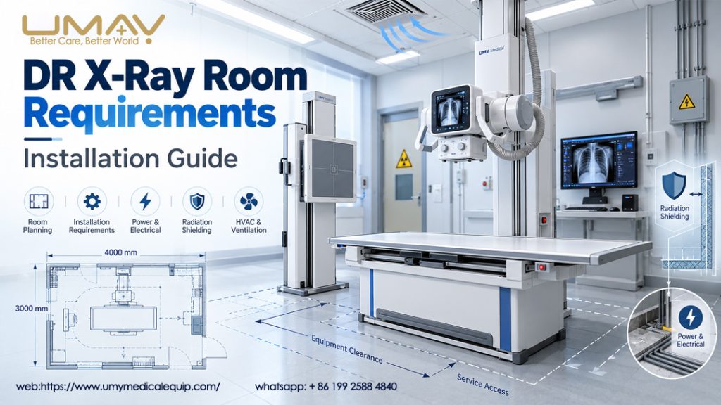
1. DR X-Ray Room Size, Layout and Equipment Clearances
Room planning begins with the type of radiography the facility will perform: standing chest examinations, table-based examinations, orthopedic imaging, or a combination. The system’s equipment footprint is only one part of the space requirement. Patients must be able to enter safely, and staff must be able to position the X-ray tube, examination table, detector stand, and accessories without obstruction.
The IAEA Safety Guide SSG-46 emphasizes facility siting, workload, patient flow, and access for stretchers as important elements of radiology-room design.
What to include in a DR room floor plan
- Patient access: door opening, wheelchair and stretcher routes, turning and transfer space.
- X-ray table: installed footprint, tabletop movement, patient approach, table loading and service clearance.
- X-ray tube support: column or rail position, tube travel, rotation envelope, and potential collision points.
- Vertical Bucky stand: detector height range and space for standing radiography.
- Operator location: shielded console, clear visibility of the patient, communication and emergency access.
- Building and maintenance: access panels, equipment delivery route, anchoring points, structural support, cable routing and cleaning access.
2. Radiation Shielding: Walls, Doors, Windows and Adjacent Areas
Radiation shielding is a professional design task, not a single material specification that can be copied between hospitals. The required barriers depend on the selected equipment, X-ray beam directions, anticipated workload, use and occupancy of adjacent areas, the geometry of the room, and local dose constraints.
According to the IAEA SSG-46 guidance, shielding should be tailored to national requirements and reassessed when equipment, procedures, workload, or adjacent occupancy changes.
| Area | What planners should verify | Who approves it |
|---|---|---|
| Primary and secondary barriers | Intended beam directions, scatter, leakage, occupancy and protective material specifications | Qualified medical physicist / radiation protection expert |
| Entrance doors | Barrier continuity, door shielding and access control | Shielding designer and local regulator |
| Control-room viewing window | Required shielding equivalence and patient observation from the console | Shielding designer |
| Ceiling and floor | Occupancy above/below and need for additional shielding | Qualified medical physicist |
| Joints and penetrations | Protection around pipes, outlets, cables, ventilation openings and wall intersections | Shielding designer / site inspector |
| Radiation signs and warnings | Applicable radiation warning symbols, access signs and warning lights | Local radiation safety authority |
For diagnostic radiography, the room design should avoid directing the primary beam toward an inadequately protected door. Operators also need a protected control position with patient visibility and communication. Shielding plans and post-installation verification may be subject to regulatory approval in the destination country.
Do not order lead-lined doors or specify wall lead equivalence solely from a generic online checklist. Request the radiation protection assessment and locally approved construction drawings first.
3. DR X-Ray Room Electrical Power, Grounding and Environment
The X-ray generator, image workstation and optional accessories can have very different power requirements. Generator output power in kW is not a direct substitute for the incoming electrical service rating in kVA or the required breaker and cable specifications.
Before ordering or installing a fixed DR system, ask the manufacturer for the electrical site-preparation documentation matching the exact generator option, including:
- Nominal supply voltage, number of phases and frequency;
- Supply capacity, permissible voltage range and transient characteristics;
- Dedicated circuits, breaker ratings, cable cross-sections and grounding/bonding requirements;
- Power-distribution and equipment connection drawings;
- UPS or controlled shutdown needs for workstation, networking and data systems;
- Ambient temperature, humidity, airflow and heat-removal requirements.
The IAEA recommends stable electrical supplies and climate control compatible with equipment manufacturers’ specified operating conditions. The detailed electrical design should be completed and checked by a qualified local engineer. Do not assume a product rated for one voltage or frequency can be installed unchanged in another market.
4. Real DR System Example: UMY Medical UMDRXY006
For an actual procurement example, UMDRXY006 Dual-Column Digital X-Ray System is a floor-mounted DR platform offered with 32 kW, 50 kW, 65 kW and 80 kW high-frequency generator configurations. It includes a movable X-ray tube column, vertical Bucky stand, four-way floating examination table, rotating-anode X-ray tube, detector options and digital imaging workstation.
| Published UMDRXY006 item | Value / configuration | Installation-planning implication |
|---|---|---|
| Generator choices | 32 / 50 / 65 / 80 kW | Select exact generator before finalizing power and tube configuration |
| 50 kW generator range | 40–150 kV; 10–630 mA | Relevant input for intended examination workload and shielding assessment |
| 50 / 65 / 80 kW electrical listing | 3-phase 380 VAC in published configuration sheet | Verify voltage, frequency and site electrical requirements for the actual order |
| Examination table | Approx. 2,000 × 730 × 630 mm | Allow additional clearance for travel, transfers and access |
| X-ray tube column height | Approx. 2,130 mm | Check ceiling and overhead service clearances |
| Tube-arm reach | Approx. 940 mm | Check motion envelope and collision clearance |
| Vertical detector stand height | Approx. 2,000 mm | Check full detector movement range and wall position |
| Detector and imaging workstation | Wired/wireless FPD options; digital imaging workstation | Confirm detector model, communication, mounting and network workflow |
These are product-page reference specifications, not approved room-construction drawings. The published 32 kW electrical specifications have multiple possible configurations, so they particularly require project-specific confirmation. The final installation manual and selected configuration take precedence over this overview.
The UMDRXY005 5.6 kW Portable Digital X-Ray Machine is a different design, with a 40–125 kV working range and 110/220 VAC or 48 V DC input options depending on configuration. Portable systems may reduce some fixed-mounting needs but do not remove the need for radiation-protection measures, suitable clinical space or local authorization. The installation and operational controls must still match the intended use and regulatory requirements.
5. Flat Panel Detectors, Workstations and PACS Connectivity
Room planning should include the digital image workflow, not only X-ray exposure hardware. A typical DR system may involve a flat panel detector, acquisition workstation, hospital network and image archive. Equipment procurement teams should confirm how the selected detector connects and how images and patient identifiers move through the system.
| Digital requirement | Question to ask before purchase |
|---|---|
| Flat panel detector | Which wired or wireless model, active area, mounting and charging/storage arrangement? |
| Acquisition workstation | Where will it sit, how will it be powered and protected, and can the operator observe the patient? |
| DICOM / PACS | Which DICOM services and integration options are included and licensed in the selected package? |
| Patient data workflow | How will examination orders, patient IDs, storage and backups be managed? |
| Network and security | Who supplies network ports, IP settings, security approvals and integration testing? |
UMDRXY006’s product information lists image acquisition, processing and dose-reporting functions, while noting that DICOM/PACS capabilities must be confirmed for the final configuration. Do not promise a specific interface or network integration until it is included in the quotation and verified in the acceptance plan.
6. Installation Sequence and Acceptance Testing
A practical DR project is best managed as a series of approvals rather than moving directly from a quotation to equipment delivery.
- Define the intended clinical workload: types of examination, expected patients, staff workflow and projected future use.
- Freeze the selected equipment configuration: generator, tube, detector, table, stands, workstation, and optional accessories.
- Obtain site-preparation documents: final footprints, travel envelopes, mounting points, electrical drawings and environmental requirements.
- Complete radiation protection design: have the qualified expert assess shielding, adjacent occupancy and applicable regulatory approvals.
- Prepare and inspect the site: construction, utilities, cabling, room finishes, IT infrastructure and access routes.
- Install and configure the system: use the manufacturer’s approved procedures and authorized service personnel.
- Carry out acceptance and commissioning tests: verify specifications, radiation safety, image quality and operating protocols under appropriate professional supervision.
- Document handover: maintain test reports, operator training records, manuals, maintenance arrangements and regulatory documentation.
IAEA SSG-46 distinguishes acceptance testing (verifying that supplied equipment meets specified requirements) from commissioning (establishing performance baselines and validating clinical-use conditions). Commissioning should be undertaken by, or under the supervision of, a qualified medical physicist before patient use.
7. DR X-Ray Room Pre-Installation Checklist for Hospital Buyers
If you are preparing a tender or requesting a DR quotation, include the information below. An accurate first inquiry helps avoid buying a system that needs unexpected electrical work, an unsuitable layout or additional accessories.
| Checklist item | Information to supply |
|---|---|
| Project and destination | Hospital/clinic, country, planned installation date, contact person |
| Room drawing | Dimensioned floor plan, ceiling height, doors/windows, adjacent occupancy, floor above/below |
| Clinical requirements | Exam types, patient volume, upright and table-based workflow |
| DR system | Preferred model and 32 / 50 / 65 / 80 kW option, or another platform |
| Detector | Required detector size, wired/wireless preference, mounting |
| Electrical | Available voltage, frequency, phases, incoming service and site engineer contact |
| Room safety | Local regulator, radiation protection consultant and status of shielding plan |
| Digital integration | PACS/RIS connection, DICOM requirements, network and IT contacts |
| Scope of supply | Table, vertical stand, collimator, accessories, workstation, printer, installation and training |
| Commercial / regulatory | Quantity, delivery address, import documents, inspection, warranty and service expectations |
A useful document package: dimensioned room plan + local electrical information + selected system configuration + intended examinations + applicable shielding assessment. These materials allow equipment and site-preparation questions to be discussed on the same basis.
8. Frequently Asked Questions
What is the minimum room size for a digital X-ray machine?
There is no universally suitable dimension. The answer depends on system geometry, full motion and clearance requirements, patient and staff access, local building standards, and the approved radiation shielding plan. Ask for the installation drawing for the exact DR model and room arrangement.
How thick should the lead shielding be in an X-ray room?
The necessary shielding cannot be determined responsibly from the X-ray generator’s kW rating alone. A qualified expert must assess intended beam directions, workload, occupancy, adjacent rooms and regulatory dose requirements before specifying protective construction.
Does a 50 kW DR X-ray system require three-phase power?
The published UMY Medical UMDRXY006 50 kW configuration lists three-phase 380 VAC, but other systems and market-specific variants may differ. Confirm the exact incoming electrical service specification with the supplier and local electrical engineer before any wiring work.
Can a hospital install a portable DR machine in any room?
No. Portability does not remove the need to assess radiation protection, access control, clinical suitability, environmental requirements and authorization under local rules. Operating conditions should be assessed for the intended location and workflow.
When can a newly installed DR X-ray system be used on patients?
Only after the applicable installation, radiation safety, acceptance, commissioning, regulatory and operational requirements have been satisfied. The facility’s medical physicist and competent local authorities should determine the required approvals and documentation.
Planning a DR X-Ray Room or Equipment Purchase?
Send UMY Medical your room dimensions or floor plan, destination country, required generator power, detector preference, and available electrical supply. We can review the available equipment configurations and quotation requirements. Final room design, shielding and approval remain the responsibility of qualified local professionals and regulators.
Technical References and Further Reading
- International Atomic Energy Agency (IAEA), SSG-46: Radiation Protection and Safety in Medical Uses of Ionizing Radiation — fixed X-ray room design, shielding and commissioning.
- World Health Organization, Portable Digital Radiography System: Technical Specifications — DR system components and procurement considerations.
- UMY Medical, UMDRXY006 32–80 kW Dual-Column Digital X-Ray System — product-specific configurations and published specifications.
- UMY Medical, UMDRXY005 5.6 kW Portable Digital X-Ray Machine — portable configuration for comparison.
Technical note: This article is a procurement and facility-planning overview, not a construction drawing, shielding calculation, electrical installation instruction, regulatory approval or substitute for an approved manufacturer installation manual.