
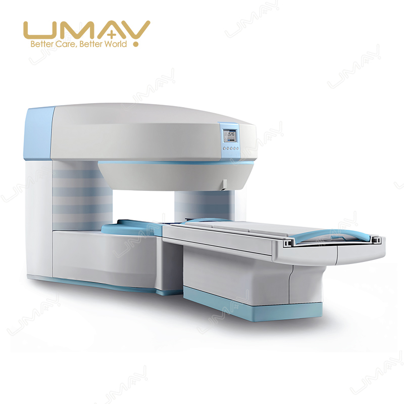
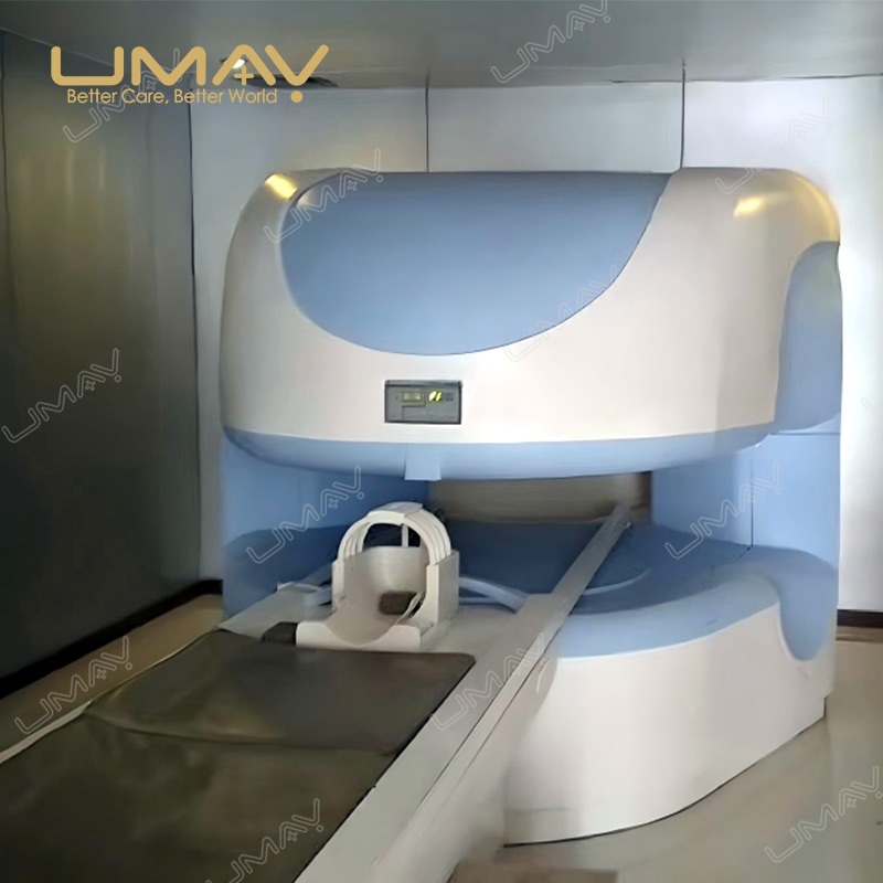
0.5T Open Permanent Magnet MRI System
The 0.5T Open Permanent Magnet MRI System is designed for hospitals and diagnostic imaging facilities that require the accessibility of an open MRI platform together with a higher permanent-magnet field strength.
The system combines a 0.5 Tesla Nd-Fe-B permanent magnet with automatic constant-temperature control, a dual-column open architecture, 25 mT/m gradient strength, 4-channel fully digital RF acquisition, phase-array receiving coils and a Windows-based MRI workstation.
Its open geometry provides a 40 cm vertical gap and wide patient access, while high-accuracy positioning, DICOM 3.0 connectivity and a comprehensive MRI sequence package support routine neurological, musculoskeletal, spinal and selected body imaging workflows.
Other MRI Field Strength Options
UMY Medical provides open permanent-magnet and superconducting MRI systems for healthcare facilities with different clinical, workflow and infrastructure requirements.
0.35T Open Permanent Magnet MRI
An open permanent-magnet MRI platform for facilities focused on routine MRI examinations and open patient access.
1.5T Superconducting MRI
A higher-field superconducting MRI platform with 16-channel digital acquisition for broader clinical imaging and higher-throughput hospital workflows.
System Highlights
0.5T Permanent Magnet
The system uses a 0.5 Tesla Nd-Fe-B permanent magnet with automatic constant-temperature control for stable magnetic-field operation.
Open Dual-Column Architecture
The rear dual-column structure provides a 40 cm vertical opening and wide horizontal accessibility, creating a more open examination environment than a conventional cylindrical MRI bore.
≤2.5 ppm Field Homogeneity
Magnetic-field homogeneity is specified at ≤2.5 ppm over a 40 cm DSV using VRMS measurement.
25 mT/m Gradient Strength
The air-cooled gradient system provides 25 mT/m single-axis gradient strength with a slew rate of 75 mT/m/ms.
4-Channel Digital RF
A fully digital transmit-and-receive RF architecture works with phase-array receiving coils for multi-channel MRI signal acquisition.
Advanced MRI Imaging Functions
The supplied software supports routine and advanced functions including FSE, STIR, FLAIR, DWI, TOF MRA, MRCP, MRU and MRM.
Open Dual-Column Design for Patient Access
The magnet uses a rear-positioned dual-column architecture designed to keep the examination area visually and physically open. Compared with a conventional cylindrical bore design, the patient has greater surrounding space and improved accessibility during positioning.
Open Examination Environment
The 40 cm vertical magnet gap and wide horizontal opening provide a less enclosed examination environment, which can be useful for patients who are uncomfortable in narrow MRI spaces.
Front and Rear Accessibility
The dual-column layout leaves the examination area more accessible from the front and rear, supporting flexible patient positioning and operator access.
Positioning Flexibility
The open structure works together with the high-accuracy patient positioning system to support examination of anatomical regions that can be more difficult to position, including shoulder and musculoskeletal applications.
0.5T Permanent Magnet System
The magnet subsystem uses Nd-Fe-B permanent-magnet technology with automatic constant-temperature regulation. Active, passive and dynamic shimming are used to support magnetic-field uniformity during MRI acquisition.
| Magnet Type | Permanent Nd-Fe-B Magnet with Automatic Constant-Temperature Control |
|---|---|
| Field Strength | 0.5T |
| Magnet Structure | Dual-Column Open Structure |
| Field Homogeneity | ≤2.5 ppm, 40 cm DSV, VRMS |
| Shim Method | Active / Passive / Dynamic |
| Vertical Gap | 40 cm |
| Horizontal Accessibility | Approx. 274° |
| 5 Gauss Fringe Field | 2.5 × 2.5 × 2.5 m |
Gradient System
The gradient subsystem provides the spatial encoding required for MRI slice selection and image acquisition. The 0.5T platform combines 25 mT/m gradient strength with a 75 mT/m/ms slew rate and an air-cooled architecture.
25 mT/m Gradient Strength
The specified single-axis gradient strength is 25 mT/m for routine and advanced MRI acquisition.
75 mT/m/ms Slew Rate
The gradient switching specification supports faster spatial encoding and multiple fast MRI sequence technologies.
Dynamic High-Order Shimming
High-order shimming can be adjusted across the X, Y and Z axes to compensate for magnetic-field variations during different examination conditions.
Air-Cooled Gradient System
The gradient coils and power electronics use air cooling, reducing the need for a dedicated gradient water-cooling circuit.
| Gradient Strength | 25 mT/m, Single Axis |
|---|---|
| Gradient Slew Rate | 75 mT/m/ms, Single Axis |
| Rise Time | 0.3 ms |
| Gradient Cooling | Air Cooled |
4-Channel Fully Digital RF System
The MRI platform uses a fully digital RF transmit-and-receive architecture with four RF acquisition channels and phase-array receiving coils.
Fully Digital Spectrometer
Digital transmit and receive architecture supports stable MRI signal control and acquisition.
4 RF Channels
Four RF channels provide multi-channel signal acquisition and support compatible phase-array receiving coils.
5 kW RF Amplifier
The RF transmitter amplifier provides a specified peak output power of 5 kW.
400 kHz Receiver Bandwidth
The receiver provides a specified bandwidth of 400 kHz per channel with a dynamic range of at least 80 dB.
| RF Architecture | Fully Digital Transmit and Receive |
|---|---|
| RF Channels | 4 Channels |
| Transmitter Peak Power | 5 kW |
| Receiver Bandwidth | 400 kHz |
| Dynamic Range | ≥80 dB |
| Noise Factor | 0.3 dB |
| Receiving Coil Type | Phase Array |
Phase-Array Receiving Coils
Dedicated phase-array receiving coils are used for different anatomical regions. The listed standard system configuration includes five main receiving-coil sets, while additional configurations can be confirmed according to the intended examinations.
| Head Coil | Available in Standard Configuration |
|---|---|
| Neck Coil | Available in Standard Configuration |
| Small Body Coil | Available in Standard Configuration |
| Large Body Coil | Available in Standard Configuration |
| Knee Coil | Available in Standard Configuration |
Other receiving-coil requirements should be confirmed according to the hospital’s examination mix before final system configuration.
Typical Clinical Applications
The 0.5T MRI platform supports routine magnetic resonance examinations across multiple anatomical regions. Final examination capability depends on the selected receiving coils, software configuration and local clinical protocols.
Neurological Imaging
MRI sequences including SE, FSE, FLAIR and DWI support routine brain and neurological imaging workflows.
Spine Imaging
The system can be configured for cervical, thoracic and lumbar spine examinations using the appropriate coil and scanning protocol.
Musculoskeletal and Joint Imaging
Dedicated receiving coils and open patient access support examinations of the knee and other selected joints and musculoskeletal structures.
Body Imaging
Body receiving coils and breath-hold imaging functions support selected abdominal and pelvic MRI examinations.
MR Angiography
TOF 2D and TOF 3D MRA functions support non-contrast magnetic resonance angiography workflows.
MRCP, MRU and MRM
Dedicated software functions support magnetic resonance cholangiopancreatography, urography and myelography applications.
Patient Table and Positioning
The patient table uses a manual positioning structure with high-accuracy positioning assistance. An isocentric positioning method helps align the selected examination region with the magnetic-field center.
This is particularly useful when positioning anatomical regions such as the shoulder, where accurate alignment with the imaging center can be more difficult.
| Patient Table | Manual Table with High-Accuracy Positioning Assistance |
|---|---|
| Maximum Patient Weight | 200 kg |
| Positioning Accuracy | ≤1 mm |
| Localizer | Included |
| Positioning Accessories | Included |
| Emergency Function | Emergency Stop / Call Function |
| Communication | Two-Way Communication System |
MRI Workstation and DICOM Connectivity
The system is supplied with a Windows-based MRI workstation for examination setup, image acquisition, image review and routine post-processing.
DICOM 3.0 and Ethernet connectivity allow the MRI system to integrate with compatible hospital imaging networks and image-management workflows.
| Workstation | MRI Workstation |
|---|---|
| Processor | Dual-Core, ≥2.8 GHz |
| Memory | ≥4 GB |
| Hard Disk | ≥1000 GB |
| Display | 24-inch LCD |
| DICOM | DICOM 3.0 |
| Network | Ethernet |
| Storage Media | CRW / DVD |
Computer hardware and workstation specifications should be reconfirmed with the final quotation because supplied computer components may change over time.
Scanning Parameters
| Maximum FOV | 400 mm |
|---|---|
| Minimum FOV | 20 mm |
| Minimum 2D Slice Thickness | 1 mm |
| Minimum 3D Slice Thickness | 0.1 mm |
| Maximum Acquisition Matrix | 1024 × 1024 |
MRI Sequences and Imaging Functions
The supplied MRI software package includes routine sequences together with multiple advanced acquisition and post-processing functions.
Routine Spin-Echo Imaging
SE 2D/3D, MSME, FSE, FDE, FRFSE, SSFSE and MSFSE are supported for routine MRI acquisition.
Inversion Recovery Imaging
IR, IRFSE, STIR and FLAIR are available for multiple tissue-contrast and fluid-sensitive imaging protocols.
Gradient-Echo Imaging
GRE 2D/3D and steady-state gradient-echo techniques are included in the imaging software package.
Diffusion-Weighted Imaging
DWI is supported with a specified maximum b-value of 1000 s/mm².
MR Angiography
TOF 2D MRA and TOF 3D MRA are supported together with MIP, MinIP and vascular image-processing functions.
MRCP, MRU and MRM
The software configuration includes MRCP, MRU and MRM functions for selected fluid-sensitive and anatomical imaging applications.
Breath-Hold Imaging
Breath-hold acquisition functions are available for selected body imaging protocols.
3D and Post-Processing
3D positioning, 3D reconstruction, multiplanar reconstruction, image filtering and online post-processing functions are included.
Standard System Configuration
The final MRI configuration should be confirmed with the quotation. The listed project configuration includes the following major system components.
- 0.5T permanent Nd-Fe-B magnet system.
- Automatic constant-temperature magnet control.
- Fully digital spectrometer.
- Preamplifier and RF amplifier.
- Flat-panel transmitter coil.
- Gradient amplifier and gradient coil.
- MRI workstation computer.
- 24-inch imaging monitor.
- Patient table and positioning accessories.
- Head receiving coil.
- Neck receiving coil.
- Small body receiving coil.
- Large body receiving coil.
- Knee receiving coil.
- Quality-control water phantom.
- MRI safety signs.
- Technical, user and maintenance documentation.
- Voltage stabilizer according to project configuration.
- Two-way patient communication system.
- Patient positioning system.
RF Shielding and Site Preparation
An RF shielding room can be included as part of the MRI project configuration. The listed shielding package can include the shielding structure, shielding door, observation window, transmission board, waveguide board, indoor lighting and an air-conditioning unit.
Civil construction, concrete foundation, floor preparation, external decoration, external electrical connection, independent grounding, equipment-room preparation and other site works should be confirmed separately before quotation and installation.
Installation, Training and Service
- Pre-installation site and utility requirement documentation.
- MRI room and equipment-room planning support.
- Equipment installation and commissioning.
- Operator training for MRI system use.
- Basic technical and maintenance training.
- Remote technical support when suitable network access is available.
Before Ordering
A 0.5T MRI installation should be configured according to the hospital’s intended examinations, expected workload, room conditions, receiving-coil requirements and local installation standards.
Please provide the following information so that UMY Medical can confirm the appropriate system and project configuration.
- Destination country and installation location.
- Required quantity.
- Expected MRI examination types.
- Expected daily patient volume.
- Required receiving-coil configuration.
- Available MRI scan-room dimensions.
- Available equipment-room dimensions.
- RF shielding-room requirements.
- Local electrical power conditions.
- Grounding and HVAC conditions.
- DICOM / PACS / RIS integration requirements.
- Required installation and training scope.
- Local registration, tender or technical documentation requirements.
If your project requires a different magnetic field strength, compare the
0.35T Open Permanent Magnet MRI System
and
1.5T Superconducting MRI System
before confirming the final MRI platform.
View more magnetic resonance imaging options in our
MRI Systems
range.
Request a 0.5T MRI Configuration and Quote
Contact UMY Medical for system configuration confirmation, project requirements, receiving-coil selection, installation planning and export quotation for the 0.5T Open Permanent Magnet MRI System.
Frequently Asked Questions
What type of magnet does the 0.5T MRI system use?
The system uses a 0.5 Tesla permanent Nd-Fe-B magnet with automatic constant-temperature control.
How open is the 0.5T MRI system?
The magnet provides a 40 cm vertical gap and approximately 274° horizontal accessibility using a dual-column open structure.
What gradient performance does the system provide?
The system provides 25 mT/m single-axis gradient strength, a 75 mT/m/ms slew rate and a 0.3 ms rise time.
Does the system support DWI, MRA and MRCP?
Yes. The listed imaging software includes DWI, TOF 2D/3D MRA, MRCP, MRU, MRM and related reconstruction and post-processing functions.
Which receiving coils are included?
The listed standard configuration includes head, neck, small body, large body and knee receiving coils. Final coil configuration should be confirmed before quotation.
What information is needed before quotation?
Please provide the destination country, intended MRI examinations, expected patient volume, required coils, available room dimensions, electrical conditions and any DICOM, installation or tender requirements.



Reviews
There are no reviews yet.