
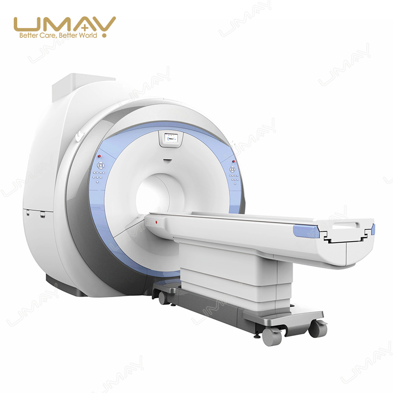
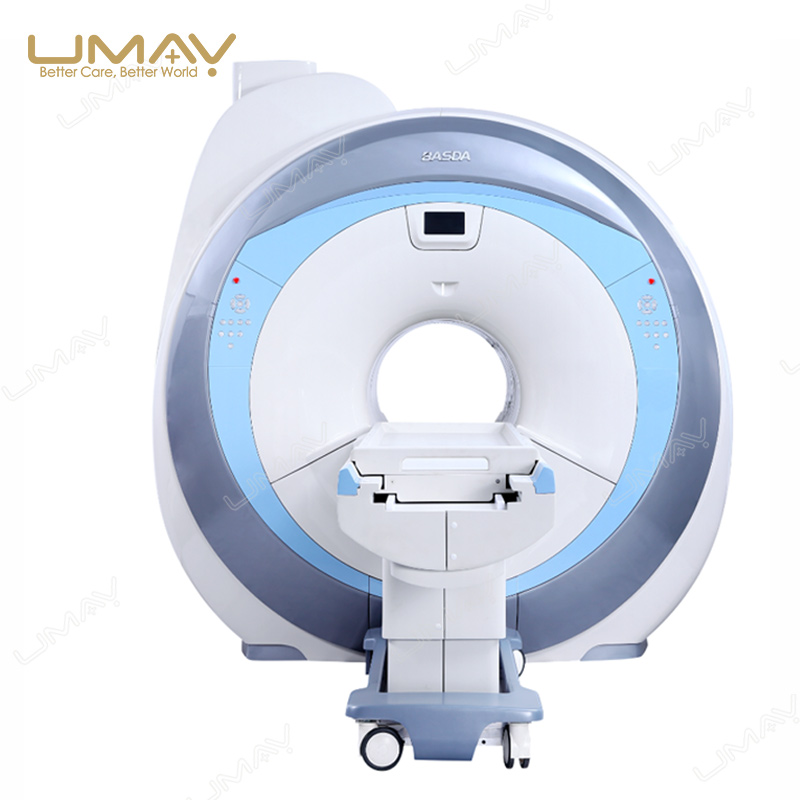
1.5T Helium-Free Superconducting MRI System
The 1.5T Helium-Free Superconducting MRI System is designed for hospitals, diagnostic imaging centers and medical projects requiring a general-purpose high-field MRI platform for neurological, body, vascular and musculoskeletal imaging.
The platform combines a 1.5 Tesla superconducting magnet with a 600 mm patient bore, 41 mT/m gradient strength, 187 mT/m/ms maximum gradient slew rate, ≥18 kW RF amplification and 16-channel fully digital parallel signal acquisition.
According to the supplied UMY specification, the magnet system has a total liquid-helium capacity of 0 L. The system also integrates a motor-driven patient table, multi-channel phased-array coils, Windows-based workstation, DICOM 3.0 connectivity and advanced MRI acquisition and post-processing technologies.
Other MRI Field Strength Options
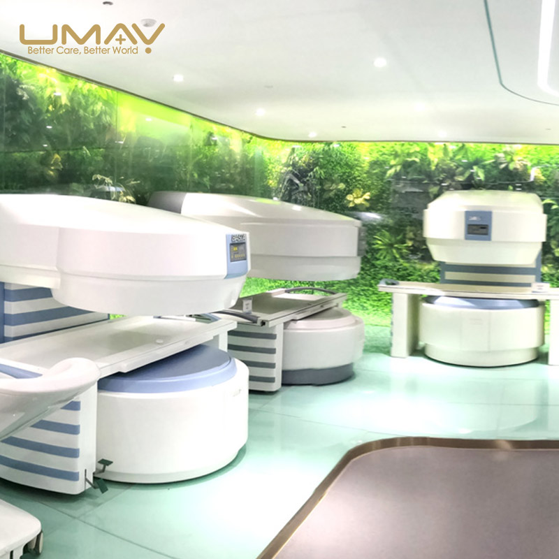
UMY Medical also supplies open permanent-magnet MRI systems for facilities with different patient-access, infrastructure and clinical requirements.
0.35T Open Permanent Magnet MRI
An open C-shaped permanent-magnet MRI platform for facilities focused on routine MRI examinations and open patient access.
0.5T Open Permanent Magnet MRI
A higher-field open permanent-magnet MRI platform with dual-column architecture, 25 mT/m gradients and 4-channel digital RF acquisition.
System Highlights
1.5T Superconducting Magnet
The superconducting magnet provides a 1.5 Tesla field with a 600 mm inner diameter and a compact magnet length of approximately 157 cm.
Helium-Free Configuration
The current UMY specification lists the total liquid-helium capacity as 0 L, distinguishing this configuration from conventional liquid-helium MRI platforms.
41 mT/m Gradient Strength
The gradient system provides up to 41 mT/m maximum gradient field strength with a maximum slew rate of 187 mT/m/ms.
16-Channel Digital RF
Sixteen parallel RF receiver channels and sixteen parallel A/D converters support multi-channel phased-array acquisition.
Advanced MRI Applications
The supplied imaging platform includes DWI, MRA, SWI, MRCP, MRU, MRM, body imaging, neuro imaging and parallel acquisition technologies.
DICOM 3.0 Workstation
A Windows-based MRI workstation with DICOM 3.0 connectivity supports image acquisition, review, storage and hospital-network integration.
1.5T Helium-Free Superconducting Magnet
The magnet is the core of the MRI system. This platform uses a 1.5T superconducting magnet with active shielding and active, passive and dynamic shimming.
The magnet has a specified stability of ≤0.1 ppm/h and provides a 600 mm inner diameter for whole-body clinical MRI examinations.
| Magnet Type | Superconductive |
|---|---|
| Field Strength | 1.5T |
| Shielding Method | Active |
| Shimming Method | Active + Passive + Dynamic |
| Magnet Stability | ≤0.1 ppm/h |
| Magnet Length | 157 cm, Excluding Cover |
| Inner Diameter | 600 mm |
| 5 Gauss Fringe Field | ≤2.5 × 2.5 × 4.0 m |
| Liquid Helium Capacity | 0 L |
Magnetic Field Homogeneity
Magnetic-field homogeneity is specified across multiple diameter spherical volumes (DSV), supporting consistent MRI signal behavior across the imaging region.
| 45 cm DSV | ≤0.832 ppm |
|---|---|
| 40 cm DSV | ≤0.323 ppm |
| 30 cm DSV | ≤0.107 ppm |
| 20 cm DSV | ≤0.041 ppm |
| 10 cm DSV | ≤0.002 ppm |
41 mT/m High-Performance Gradient System
The gradient subsystem determines spatial encoding performance and contributes directly to fast imaging, thin-slice acquisition and advanced MRI sequence capability.
41 mT/m Maximum Gradient Field
The system provides a maximum gradient field strength of 41 mT/m.
187 mT/m/ms Slew Rate
The specified maximum gradient slew rate is 187 mT/m/ms, with a minimum gradient rise time of 0.22 ms.
Simultaneous Maximum Performance
The specification states that maximum gradient strength and maximum slew rate can be reached at the same time.
Water-Cooled Gradient System
The gradient subsystem uses water cooling for thermal management during clinical scanning.
| Maximum Gradient Field | 41 mT/m |
|---|---|
| Maximum Gradient Slew Rate | 187 mT/m/ms |
| Minimum Rise Time | 0.22 ms |
| Cooling System | Water Cooling |
| Gradient Control | Full-Digital Real-Time Control |
16-Channel Fully Digital RF System
The RF subsystem uses fully digital transmission and reception with sixteen parallel receiving channels and sixteen parallel A/D converters.
≥18 kW RF Amplifier
The RF subsystem provides a specified maximum amplifier power of at least 18 kW.
16 Parallel Receiver Channels
Sixteen parallel RF receiving channels support multi-channel phased-array signal acquisition.
16 Parallel A/D Converters
Each parallel acquisition channel is supported by dedicated digital signal conversion for MRI data acquisition.
Parallel Acquisition Platform
The system supports parallel acquisition technology and multi-channel phased-array receiving coils.
| RF System | Fully Digital |
|---|---|
| RF Amplifier Power | ≥18 kW |
| Center Frequency | 63.87 MHz |
| Receiver Channels | 16 |
| Parallel A/D Converters | 16 |
| Receiver Bandwidth per Channel | ≥1.0 MHz |
| Transmission Bandwidth | 550 kHz |
| Receiver Signal Resolution | ≥16 bit |
| RF Receiver Noise Level | ≤0.45 dB |
| Coil Type | Multi-Channel Phased Array |
MRI Coil Configuration
The standard Helium-Free MRI configuration includes anatomical and flexible receiving coils for routine clinical examinations.
| Head Coil | Standard Configuration |
|---|---|
| Neck Coil | Standard Configuration |
| Body Coil | Standard Configuration |
| Knee Coil | Standard Configuration |
| Flexible Coil | Standard Configuration |
| Respiratory Gating | Standard Configuration |
Additional coil or gating requirements should be confirmed according to the intended examination mix before quotation.
Typical Clinical Applications
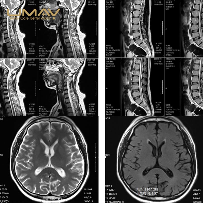
The 1.5T MRI platform supports a broad range of clinical imaging applications through its supplied MRI sequence library, phased-array coils and advanced imaging software.
Neurological Imaging
Neuro imaging functions, DWI, FLAIR, SWI and other MRI sequences support routine brain and neurological examinations.
Spine Imaging
The system supports spinal MRI examinations and includes MR myelography functions for selected clinical applications.
Body Imaging
Body imaging, phase/de-phase techniques, breath-related acquisition functions and fat-suppression sequences support abdominal and other selected body examinations.
MR Angiography
MRA capability includes 2D and 3D TOF imaging, continuous multi-layer 3D TOF, MRA cropping and maximum intensity projection.
MRCP, MRU and MRM
The supplied advanced imaging package includes magnetic resonance cholangiopancreatography, urography and myelography.
Musculoskeletal Imaging
Knee and flexible coils support joint and musculoskeletal MRI examinations according to the selected protocol and coil configuration.
Advanced MRI Sequences and Imaging Technology
Spin-Echo Imaging
SE 2D/3D, FSE 2D/3D and single-shot FSE are included for routine anatomical imaging.
Fat and Water Suppression
The system includes spin-echo fat suppression, frequency-selective fat suppression, water suppression, FIR, FLAIR and STIR.
Diffusion-Weighted Imaging
DWI is included, with the current specification listing a maximum b-value of 10000.
Susceptibility-Weighted Imaging
SWI is included in the advanced imaging technology package.
Parallel Acquisition
Parallel acquisition supports both image-based and K-space-based algorithms with a specified maximum acceleration factor of 4.
Artifact Correction
The software includes fluid compensation, respiratory compensation, head-motion artifact correction, eddy-current correction and gradient-linearity correction.
Motor-Driven Patient Table and Scan Environment
The system uses a motor-driven patient table with positioning accuracy of ≤1 mm. Feet-first entry, emergency table control and controls on both sides of the scanner are included in the supplied specification.
| Patient Table | Motor-Driven |
|---|---|
| Positioning Accuracy | ≤1 mm |
| Feet-First Entry | Supported |
| Emergency Table Control | Supported |
| Scanner-Side Table Controls | Both Sides |
| Lighting / Ventilation / Call System | Adjustable |
MRI Workstation and DICOM Connectivity
The MRI workstation uses UMY MRI software on a Windows platform and supports examination setup, image acquisition, image review and hospital imaging-network integration.
| MRI Software | UMY MRI Software |
|---|---|
| Operating System | Windows 10 |
| CPU | ≥3.0 GHz |
| Memory | ≥8 GB |
| Storage | ≥1000 GB |
| Display | 24-inch LCD |
| Monitor Resolution | 1920 × 1200 |
| External Storage | DVD / USB |
| DICOM | DICOM 3.0 |
| Fiber Technology | Available |
Scanning Parameters
| Minimum 2D Slice Thickness | 0.1 mm |
|---|---|
| Minimum 3D Slice Thickness | 0.05 mm |
| Maximum FOV | 50 cm |
| Minimum FOV | 1 cm |
| Parallel Acquisition Acceleration Factor | Up to 4 |
Standard System Configuration
The final configuration should be confirmed with the quotation. The current Helium-Free MRI configuration includes the following major components.
- 1.5T Helium-Free superconducting magnet system.
- Gradient system.
- Full-digital spectrometer.
- RF amplifier.
- RF coil system.
- Scan console and PC system.
- 24-inch IPS LCD display.
- Operator desk.
- Head coil.
- Neck coil.
- Body coil.
- Knee coil.
- Flexible coil.
- Respiratory gating system.
- Electric patient table.
- Positioning accessories.
- Quality-control set.
- Cooling chiller.
- System signal-processing device.
- Power-control system.
- Quench device.
- Two-way voice and patient-call system.
- Excitation power cabinet.
- UMY V70 imaging software.
RF Shielding and Site Preparation
The project configuration can include an RF shielding room with shielding structure, shielding door, observation window, transmission board, waveguide board, indoor lighting and an indoor air-conditioning unit.
Civil construction, external shielding-room decoration, concrete foundation, floor preparation, operating room, equipment room, external air conditioning, external electrical connection and independent grounding are listed as hospital responsibilities in the supplied project configuration and should be confirmed before quotation.
1.5T MRI or Open Permanent-Magnet MRI?
MRI system selection should be based on the intended examination mix, expected patient volume, site conditions, infrastructure requirements and future clinical development rather than field strength alone.
Consider 1.5T When
The project requires a general-purpose high-field MRI platform, broader advanced imaging functions, multi-channel acquisition and higher clinical throughput potential.
Consider 0.5T When
Open patient access remains a major requirement, while a higher-field permanent-magnet platform is preferred over a 0.35T system.
Consider 0.35T When
The primary requirement is routine MRI in an open permanent-magnet architecture with a comparatively simpler system concept.
Before Ordering
A 1.5T MRI project requires coordinated planning of the scanner, coils, software, shielding, electrical supply, cooling and installation environment.
Please provide the following information so that the final MRI configuration and quotation can be reviewed.
- Destination country and installation location.
- Required quantity.
- Expected MRI examination types.
- Expected daily patient volume.
- Required receiving-coil configuration.
- Required clinical software and imaging packages.
- Available MRI scan-room dimensions.
- Available equipment-room and control-room dimensions.
- RF shielding requirements.
- Local electrical power conditions.
- Cooling and HVAC conditions.
- Grounding requirements.
- DICOM / PACS / RIS integration requirements.
- Installation and training requirements.
- Local registration, tender or technical documentation requirements.
If an open permanent-magnet MRI is more appropriate for the project, compare the
0.5T Open Permanent Magnet MRI System or 0.35T Open Permanent Magnet MRI System.
Request a 1.5T MRI Configuration and Quote
Contact UMY Medical to confirm the Helium-Free MRI configuration, receiving coils, software packages, site requirements, installation scope and export quotation.
Frequently Asked Questions
What is a 1.5T Helium-Free MRI system?
It is a 1.5 Tesla superconducting MRI system whose supplied UMY specification lists a total liquid-helium capacity of 0 L.
What is the bore diameter of the 1.5T MRI system?
The superconducting magnet has a 600 mm inner diameter.
What is the gradient performance?
The system provides a maximum gradient field of 41 mT/m, a maximum slew rate of 187 mT/m/ms and a minimum rise time of 0.22 ms.
How many RF receiver channels are available?
The RF system provides 16 parallel receiver channels together with 16 parallel A/D converters.
Does the system support DWI, MRA and SWI?
Yes. The supplied software specification includes DWI, 2D/3D TOF MRA, SWI, MTC and maximum-intensity-projection functions.
Does the system support MRCP, MRU and MRM?
Yes. MRCP, MRU and MRM are included in the listed advanced imaging technology package.
Which coils are included in the standard configuration?
The current Helium-Free configuration lists head, neck, body, knee and flexible coils, together with respiratory gating.
Does the system support DICOM connectivity?
Yes. The workstation specification includes DICOM 3.0 and a Windows-based MRI operating platform.
What should be confirmed before purchasing a 1.5T MRI system?
The final project should confirm the examination mix, coils, software packages, MRI-room dimensions, RF shielding, electrical supply, cooling, grounding, DICOM integration, installation responsibilities and local documentation requirements.



Reviews
There are no reviews yet.