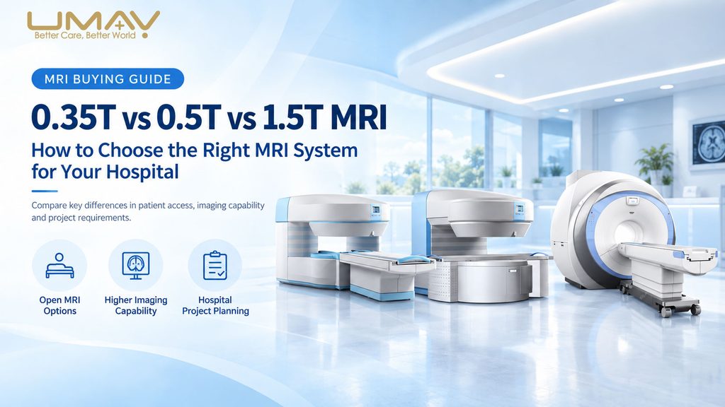
Choosing between a 0.35T, 0.5T and 1.5T MRI system is not simply a matter of selecting the highest magnetic field strength. Hospitals and diagnostic imaging centers should evaluate the expected examination mix, patient volume, patient-access requirements, gradient performance, RF channels, receiving coils, MRI software, installation conditions and long-term clinical development before selecting a system.
For UMY Medical’s current MRI range, the 0.35T system is positioned as an open permanent-magnet platform for routine MRI applications, the 0.5T system provides a higher-field open permanent-magnet platform with more clearly defined gradient and advanced imaging capabilities, and the 1.5T system is a helium-free superconducting MRI platform designed for broader general-purpose and advanced clinical imaging.
0.35T vs 0.5T vs 1.5T MRI: Quick Comparison
| Comparison | 0.35T Open MRI | 0.5T Open MRI | 1.5T MRI |
|---|---|---|---|
| Magnet Type | Permanent Nd-Fe-B | Permanent Nd-Fe-B | Superconducting |
| Scanner Design | Open C-shaped | Open dual-column | 600 mm bore |
| Magnetic Field Strength | 0.35T | 0.5T | 1.5T |
| RF Acquisition | 4-channel digital | 4-channel fully digital | 16-channel fully digital |
| Gradient Strength | Confirm with final configuration | 25 mT/m | 41 mT/m |
| Gradient Slew Rate | Confirm with final configuration | 75 mT/m/ms | 187 mT/m/ms |
| Patient Access | Open C-shaped structure | 40 cm vertical gap with wide open access | 600 mm cylindrical bore |
| Patient Table | Manual | Manual with high-accuracy positioning assistance | Motor-driven |
| Clinical Positioning | Routine open MRI | Higher-field open MRI | General-purpose high-field MRI |
| Typical Imaging Scope | Routine neuro, spine, joint and selected body imaging | Routine imaging plus listed DWI, MRA, MRCP, MRU and MRM functions | Broader neuro, body, vascular and musculoskeletal imaging |
| Liquid-Helium Magnet System | No | No | Current UMY helium-free configuration: 0 L specified liquid-helium capacity |
Does Higher MRI Field Strength Always Mean Better MRI?
No. Magnetic field strength is important, but it is only one component of MRI performance.
Higher field strength can provide more available MR signal, which can be used to support higher spatial resolution, shorter acquisition times or more demanding imaging techniques. However, the final imaging capability of an MRI scanner also depends on the gradient system, RF acquisition channels, receiving coils, pulse sequences, reconstruction software, patient positioning and protocol design.
This is why two MRI systems with the same Tesla rating may still differ substantially in clinical capability and workflow.
For hospital procurement, the more useful question is not simply “Which MRI has the highest Tesla?” but “Which complete MRI configuration matches our examinations, patient volume, infrastructure and future clinical requirements?”
Field Strength Is Only One Part of MRI Performance
| MRI Subsystem | Why It Matters |
|---|---|
| Magnet | Provides the static magnetic field and influences available MR signal. |
| Gradient System | Controls spatial encoding, slice selection and many fast imaging capabilities. |
| RF System | Transmits RF energy and receives MR signals from the patient. |
| Receiving Coils | Determine which anatomical regions can be examined efficiently and influence local signal reception. |
| Sequences | Determine which tissue contrasts and clinical imaging techniques are available. |
| Reconstruction and Software | Provide image processing, artifact correction, advanced imaging and workflow functions. |
Hospitals should therefore compare complete MRI configurations rather than Tesla values alone.
When Does a 0.35T Open MRI Make Sense?
A 0.35T open MRI can be considered when the primary goal is to establish routine MRI capability in an open permanent-magnet architecture.
The UMY 0.35T MRI uses a C-shaped Nd-Fe-B permanent magnet with automatic constant-temperature control and an all-digital 4-channel RF acquisition system. It supports configurable anatomical receiving coils and uses a manually operated patient table.
With the appropriate coils and installed software, the system can be configured for routine examinations including brain and head MRI, cervical and spine imaging, knee and other joint imaging, shoulder imaging, wrist and ankle imaging and selected body examinations.
The 0.35T platform therefore makes the most sense when open patient access and routine MRI coverage are more important than obtaining the broader advanced imaging configuration of a higher-field superconducting system.
One important procurement point is that the currently available 0.35T documentation does not specify the gradient subsystem in the same detail as the 0.5T and 1.5T products. Gradient performance and required advanced software should therefore be confirmed in the final quotation rather than assumed.
View the 0.35T Open Permanent Magnet MRI System →
Why Is 0.5T MRI an Important Middle Option?
The 0.5T MRI occupies a useful position between a lower-field open MRI and a 1.5T superconducting MRI.
It retains an open permanent-magnet architecture while increasing magnetic field strength and providing a more clearly specified gradient and imaging platform.
The UMY 0.5T system uses a dual-column Nd-Fe-B permanent magnet with automatic constant-temperature control. The specification lists a 40 cm vertical gap, approximately 274° horizontal accessibility and field homogeneity of ≤2.5 ppm over a 40 cm DSV.
Its gradient system provides 25 mT/m single-axis gradient strength, a 75 mT/m/ms slew rate and a 0.3 ms rise time. The RF platform uses four fully digital transmit-and-receive channels with phase-array receiving coils.
The listed software package includes SE, GRE, FSE, STIR, FLAIR, DWI, TOF MRA, MRCP, MRU and MRM functions.
For a hospital that wants to preserve the advantages of an open examination environment while requiring a stronger and more clearly defined permanent-magnet MRI platform, 0.5T can therefore be a distinct project category rather than simply a compromise between 0.35T and 1.5T.
View the 0.5T Open Permanent Magnet MRI System →
When Is a 1.5T MRI the More Appropriate Choice?
A 1.5T MRI is positioned for hospitals and diagnostic imaging centers requiring a broader general-purpose clinical platform, stronger gradient performance, more RF receiving channels and more advanced imaging capabilities.
The current UMY 1.5T helium-free configuration uses a superconducting magnet with a 600 mm inner diameter and a magnet length of approximately 157 cm.
The gradient system provides up to 41 mT/m maximum gradient strength and 187 mT/m/ms maximum slew rate with a minimum specified rise time of 0.22 ms.
The RF subsystem provides ≥18 kW amplifier power, 16 parallel receiver channels, 16 parallel A/D converters and multi-channel phased-array coil support.
The imaging software includes neurological and body imaging, DWI, 2D/3D TOF MRA, SWI, MRCP, MRU, MRM, parallel acquisition and multiple artifact-correction technologies.
For hospitals planning a broad examination mix or a larger MRI service, these hardware and software capabilities may be more important than open magnet geometry.
View the 1.5T Helium-Free Superconducting MRI System →
Open MRI vs Superconducting MRI
The difference between open permanent-magnet MRI and superconducting MRI involves both scanner geometry and system architecture.
Open Permanent-Magnet MRI
The UMY 0.35T and 0.5T scanners use permanent Nd-Fe-B magnets and open examination structures.
The 0.35T system uses a C-shaped magnet, while the 0.5T system uses a dual-column open architecture. These designs provide greater physical access around the patient than a conventional cylindrical MRI bore.
Permanent-magnet systems also do not use a superconducting liquid-helium magnet system.
1.5T Superconducting MRI
The UMY 1.5T system uses a superconducting magnet with a 600 mm cylindrical patient bore. Its clinical positioning is based on the combination of higher field strength, stronger gradients, 16-channel digital acquisition and a broader advanced imaging package.
Its site-planning requirements are different from those of a permanent-magnet MRI and should be evaluated using the final project-specific installation documentation.
Helium-Free 1.5T MRI vs Traditional Liquid-Helium MRI
Traditional superconducting MRI systems have historically relied on cryogenic magnet systems containing liquid helium. MRI procurement therefore often includes consideration of cryogenic hardware, cooling systems, magnet monitoring and service requirements.
The current UMY 1.5T helium-free specification lists the magnet as superconductive while specifying a total liquid-helium capacity of 0 L.
This distinction is important when comparing 1.5T MRI proposals because the term “1.5T MRI” alone does not describe the magnet’s cryogenic architecture.
Hospitals should confirm the exact magnet configuration, cooling system, quench-management design, maintenance requirements and site responsibilities in the final technical proposal rather than assuming that all 1.5T systems use the same superconducting architecture.
How Do Clinical Applications Differ?
| Clinical Requirement | 0.35T | 0.5T | 1.5T |
|---|---|---|---|
| Routine Brain MRI | Supported with appropriate configuration | Supported | Supported |
| Spine Imaging | Supported with appropriate configuration | Supported | Supported |
| Joint / Musculoskeletal MRI | Supported | Supported | Supported |
| Selected Body Imaging | Configuration dependent | Supported with appropriate coils and protocols | Broader body imaging package |
| DWI | Confirm final software package | Listed | Listed |
| TOF MRA | Confirm final software package | 2D / 3D listed | 2D / 3D listed |
| MRCP | Confirm final software package | Listed | Listed |
| SWI | Confirm final software package | Not listed in current supplied specification | Listed |
| Parallel Acquisition | Confirm final configuration | Confirm final configuration | Listed, acceleration factor up to 4 |
The table should not be interpreted as a substitute for final protocol validation. The exact clinical capability depends on the installed coil, software and sequence configuration supplied with the system.
Why Coils and MRI Software Matter as Much as Field Strength
A frequent purchasing mistake is to compare MRI scanners using field strength alone while treating coils and software as secondary accessories.
In practice, the receiving-coil package directly affects which anatomical regions the hospital can examine efficiently, while the software package determines whether advanced functions such as DWI, MRA, SWI, MRCP or parallel acquisition are available.
For example, the current UMY 0.35T documentation lists multiple anatomical coil options but advises that the final software and advanced applications should be confirmed according to the project configuration.
The 0.5T documentation provides a more explicitly defined sequence package, while the 1.5T helium-free configuration provides an even broader advanced imaging set.
Hospitals should therefore request a final quotation that lists the exact receiving coils, software licenses, clinical packages and optional applications instead of purchasing only by scanner model.
How Does MRI Field Strength Affect Patient Throughput?
Patient throughput cannot be determined from field strength alone.
MRI workflow is influenced by sequence design, protocol length, patient preparation, coil changes, patient positioning, operator experience, reconstruction speed and the complexity of the requested examination.
Higher-performance gradients, more RF channels and parallel acquisition can support faster or more flexible workflows, but actual patients-per-day capacity should be calculated from the hospital’s own examination mix.
For this reason, procurement teams should avoid comparing MRI systems using a single advertised scan-time figure unless the underlying protocol and examination conditions are clearly defined.
What Site Requirements Should Be Checked Before Buying MRI?
MRI procurement is a site-planning project as well as an equipment purchase.
Before selecting the system, the hospital should confirm scan-room dimensions, equipment-room and control-room requirements, delivery access, RF shielding, electrical supply, grounding, HVAC, cooling requirements, magnetic fringe-field planning and the responsibilities of the scanner supplier and hospital.
The 0.5T project documentation, for example, includes an RF shielding-room option but separately identifies civil works, foundation, external electrical connections, grounding and other site work that must be confirmed by the customer.
The 1.5T helium-free configuration also includes an RF shielding option and requires its own cooling, electrical and superconducting-magnet site planning.
Final room dimensions and utilities should always follow the site-planning document for the selected MRI configuration rather than a generic online room-size recommendation.
Does a Small Hospital Really Need a 1.5T MRI?
Not necessarily.
A smaller hospital should first identify the examinations it intends to provide, its expected daily patient volume, referral profile, specialist services and future imaging plans.
A facility mainly performing routine brain, spine and musculoskeletal MRI may find that an open permanent-magnet platform covers its planned service requirements.
A hospital planning a broader mix of neurological, vascular, body and advanced imaging may obtain more value from the gradient, RF and software capabilities of a 1.5T platform.
Hospital size alone should therefore not determine MRI field strength.
What Is the Total Cost of an MRI Project?
The purchase price of the scanner is only one part of MRI project cost.
Hospitals should also evaluate room preparation, RF shielding, power distribution, grounding, HVAC or cooling equipment, receiving coils, software options, installation, commissioning, training, preventive maintenance, replacement parts and potential equipment downtime.
The exact cost structure differs significantly between permanent-magnet and superconducting MRI projects, so quotations should clearly separate scanner hardware from site work and long-term service responsibilities.
A lower initial scanner price does not automatically mean a lower total project cost, and a higher-cost scanner does not automatically provide better value if its additional capabilities are not required by the hospital.
Which MRI System Fits Different Hospital Requirements?
| Project Requirement | MRI Platform to Evaluate |
|---|---|
| Routine MRI with open C-shaped patient access | 0.35T Open Permanent Magnet MRI |
| Open MRI with a higher permanent-magnet field strength | 0.5T Open Permanent Magnet MRI |
| Open MRI requiring listed DWI, MRA and MRCP capabilities | 0.5T Open Permanent Magnet MRI |
| General-purpose MRI for a broader hospital examination mix | 1.5T Helium-Free Superconducting MRI |
| 16-channel RF and stronger gradient performance | 1.5T Helium-Free Superconducting MRI |
| Advanced neuro, vascular, SWI and broader body imaging | 1.5T Helium-Free Superconducting MRI |
These are project-selection directions rather than universal rules. Final selection should be based on the hospital’s examination requirements, site conditions and confirmed product configuration.
What Should Be Included in an MRI RFQ?
- Destination country and installation location.
- Expected MRI examination types.
- Expected daily patient volume.
- Required field strength and preferred scanner geometry.
- Required receiving coils.
- Required DWI, MRA, SWI, MRCP, MRU, MRM or other software packages.
- Available scan-room, equipment-room and control-room dimensions.
- RF shielding-room status.
- Local electrical supply and grounding conditions.
- HVAC and cooling conditions.
- DICOM, PACS and RIS integration requirements.
- Required installation and commissioning scope.
- Operator and engineer training requirements.
- Required warranty and maintenance scope.
- Local registration, tender and technical-document requirements.
What Should Be Checked Before MRI Project Handover?
Before final acceptance, the hospital should confirm that the delivered scanner configuration matches the purchase contract and technical proposal.
The final check should include the magnet and gradient configuration, supplied RF coils, installed MRI software, licensed clinical packages, workstation, DICOM connectivity, patient table, communication systems and any shielding or cooling equipment included in the contract.
The hospital should also confirm installation and commissioning records, operator training, basic maintenance documentation, technical manuals and the agreed after-sales support process.
For tender and distributor projects, documenting the final delivered configuration is particularly important because MRI performance depends on the complete system rather than the scanner cabinet alone.
Frequently Asked Questions
What is the main difference between 0.35T, 0.5T and 1.5T MRI?
The main differences include magnet technology, field strength, scanner geometry, gradient performance, RF acquisition channels and available clinical software. The UMY 0.35T and 0.5T systems use open permanent magnets, while the current 1.5T configuration uses a helium-free superconducting magnet.
Is 1.5T MRI always better than 0.5T MRI?
No. A 1.5T system provides a higher-field general-purpose platform with stronger gradients and more RF channels, but an open 0.5T system may be more appropriate when open patient access and permanent-magnet architecture are important project requirements.
What is the difference between 0.35T and 0.5T open MRI?
Both systems use open permanent magnets and 4-channel digital RF acquisition. The 0.5T system provides a higher magnetic field strength and has more detailed specified gradient and advanced imaging capabilities, including 25 mT/m gradients, 75 mT/m/ms slew rate and listed DWI, MRA and MRCP functions.
Does open MRI use liquid helium?
The UMY 0.35T and 0.5T systems use Nd-Fe-B permanent magnets and do not use a superconducting liquid-helium magnet system.
Is the UMY 1.5T MRI helium-free?
The current UMY helium-free 1.5T specification lists a superconducting magnet with a total liquid-helium capacity of 0 L.
How many RF channels does each MRI system use?
The current UMY 0.35T and 0.5T configurations use 4-channel digital RF acquisition. The 1.5T helium-free system uses 16 parallel RF receiving channels and 16 parallel A/D converters.
Which MRI supports DWI and MRA?
The current 0.5T and 1.5T specifications explicitly list DWI and MRA functions. Advanced imaging availability for the 0.35T system should be confirmed according to the final software configuration.
Which MRI is suitable for a small hospital?
The choice depends on the planned examination mix, patient volume, infrastructure, required software and future clinical development. A small hospital does not automatically need a 1.5T system, but a lower-field MRI is also not automatically sufficient for every project.
What should a hospital confirm before purchasing MRI?
The hospital should confirm field strength, gradient performance, RF channels, receiving coils, imaging software, expected patient volume, room conditions, RF shielding, power, cooling, DICOM integration, installation scope, training and long-term service requirements.
Compare UMY MRI Systems
UMY Medical currently provides open permanent-magnet and helium-free superconducting MRI configurations for different hospital and diagnostic imaging requirements.
0.35T Open Permanent Magnet MRI System →
0.5T Open Permanent Magnet MRI System →
1.5T Helium-Free Superconducting MRI System →
For additional MRI project information, visit our MRI Systems page or contact UMY Medical with your expected examinations, patient volume and site conditions.