
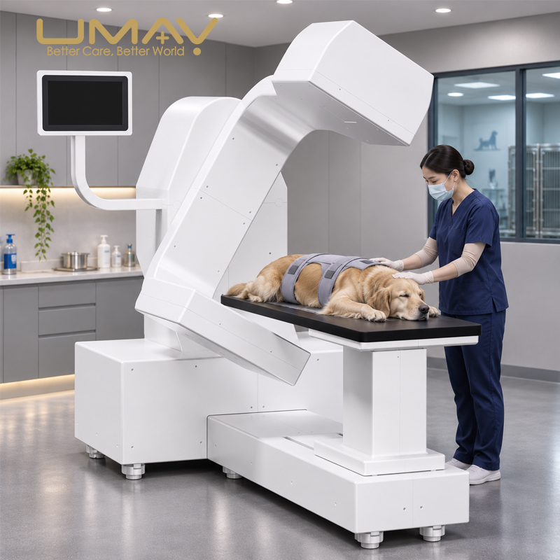
UMH-JH01B Open-C-Arm Veterinary CT Scanner
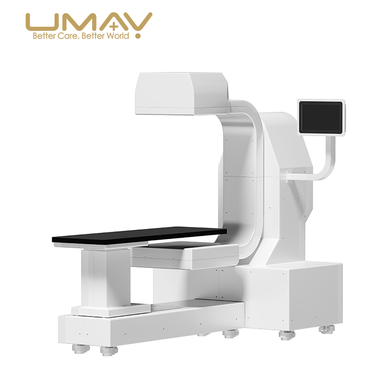
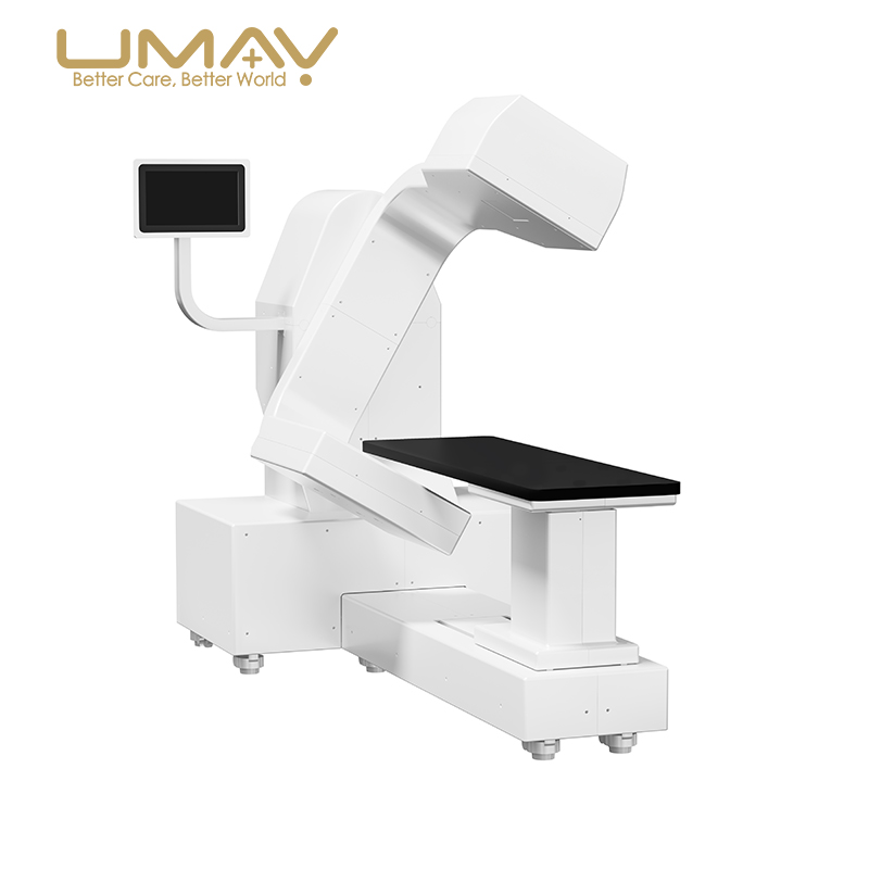
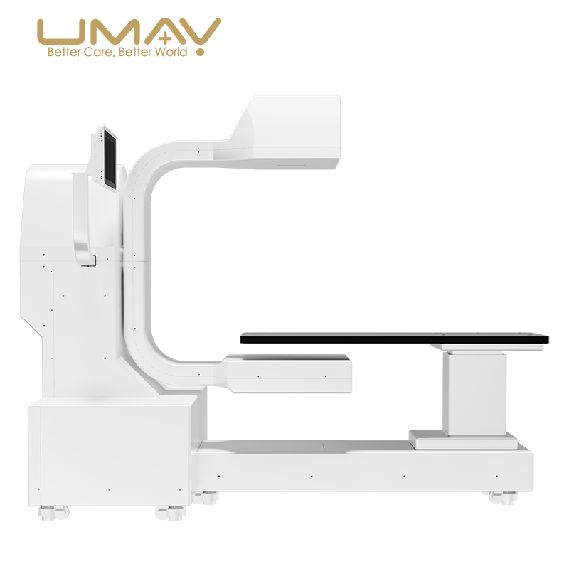
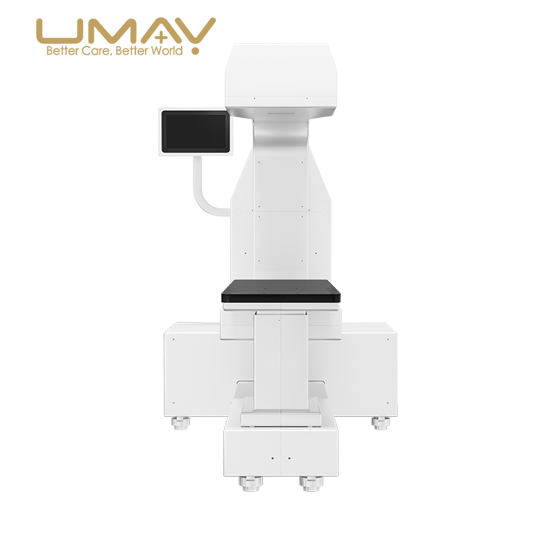
The UMH-JH01B Open-C-Arm Veterinary CT Scanner is a compact computed tomography imaging system intended for veterinary clinics and animal hospitals. Its open C-arm structure supports access to the examination area for animal positioning, while the compact mechanical layout is designed for facilities with limited space.
The manufacturer lists a 15 kW X-ray tube, 3072 × 3072 detector resolution, 0.14–0.50 mm CT slice thickness and minimum scan time of 3.5 seconds. The system also incorporates adjustable patient-table positioning and a left-right adjustable display.
Key Features
- Open C-arm architecture: Open access to the examination area to support animal positioning.
- Space-conscious configuration: Designed for veterinary facilities seeking a compact CT installation footprint.
- Adjustable operator display: Left-right screen adjustment supports flexible equipment layout.
- Three-segment scanning and stitching: Manufacturer-described function for combining scan segments.
- Respiratory gating synchronization: Listed as a function intended to help manage respiratory-motion effects.
- Intelligent three-phase contrast peak tracking: Manufacturer-listed contrast timing feature.
- Multiple acquisition settings: Brochure lists nine scan modes and three image-quality grades, without providing their individual names.
- 3D printing workflow: Brochure describes connecting imaging visualization to 3D printed models for preoperative planning; compatible equipment and workflow should be confirmed.
Technical Specifications
Detector and Imaging
| Detector | O-S (manufacturer designation; confirm specification) |
|---|---|
| Detector resolution | 3072 × 3072 |
| X-ray tube power | 15 kW |
| X-ray tube heat capacity | 150 kJ |
| CT slice thickness | 0.14–0.50 mm |
| Maximum field of view | 270 × 580 mm (manufacturer-listed) |
| Number of scan layers | 550–1200 (manufacturer-listed) |
| Minimum scan time | 3.5 seconds |
Table and Dimensions
| Table travel range | 360 mm |
|---|---|
| Table elevation range | 295 mm |
| Approximate overall dimensions (L × W × H) | 2215 × 1100 × 1730 mm |
These figures are transcribed from the supplied product brochure and are for reference. Final dimensions, detector designation, installation requirements and delivered configuration must be confirmed by the supplier before purchase.
Veterinary CT Imaging and Workflow
Open Access and Animal Positioning
The open C-arm design is intended to simplify access to the examination table for veterinary patient setup. Table travel of 360 mm and elevation adjustment of 295 mm are listed in the brochure. Appropriate patient positioning and immobilization remain clinical decisions.
Scanning, Stitching and Contrast Features
The brochure highlights three-segment scanning and stitching, respiratory gating synchronization, and intelligent three-phase contrast peak tracking. It also states that nine scan modes and three image-quality grades are available. Confirm detailed protocols, contrast-injection requirements and the installed software configuration before procurement.
Visualization and 3D Printing
The manufacturer describes a workflow connecting CT visualization with 3D printing to support the creation of tangible anatomical models and preoperative simulation. Printer models, data formats and third-party integrations are not specified in the supplied brochure and should be verified separately.
Typical Applications
- CT imaging services in veterinary clinics and animal hospitals.
- Diagnostic imaging and preoperative planning for dogs, cats and exotic animal patients, as described in the brochure.
- Veterinary facilities considering an open-access CT system for space-constrained imaging rooms.
Installation and Purchasing Considerations
Before requesting a quotation, provide the destination country, available room dimensions, intended animal types, expected clinical applications, required image-processing functions and any existing workstation or hospital information systems.
The supplied brochure does not specify electrical input requirements, radiation shielding specifications, total system weight, anesthesia provisions, included workstation hardware, DICOM interoperability, regulatory certifications or standard accessory scope. Obtain configuration-specific documentation and confirm applicable local requirements before installation.
Frequently Asked Questions
Is the UMH-JH01B a conventional closed-ring CT scanner?
No. The supplied brochure identifies the UMH-JH01B as an open C-arm veterinary CT system, designed to provide easier access during animal positioning.
What CT slice thickness does the UMH-JH01B support?
The manufacturer lists a CT slice thickness range of 0.14–0.50 mm. Confirm the acquisition protocol associated with the required setting.
Does the system support respiratory gating and image stitching?
Yes. Both respiratory gating synchronization and three-segment scanning and stitching are described in the brochure. Configuration and clinical workflow details should be confirmed with the supplier.
Can veterinary CT data be used for 3D printed models?
The brochure describes a connection to 3D printing workflows for anatomical models. Hardware, software licenses and compatible export formats are not specified.
What details are needed to request a quotation?
Please share the destination country, quantity, planned veterinary applications, room dimensions, required scan protocols and any workstation or 3D printing requirements.
Related Veterinary and Radiological Equipment
Explore other solutions in the UMY Medical Veterinary Equipment and Radiological Equipment collections. Check each model’s specifications to select the appropriate imaging technology for your facility.
Request a Quote for UMH-JH01B
Contact UMY Medical with your veterinary CT requirements, destination country and planned installation details for configuration review and pricing.


Reviews
There are no reviews yet.