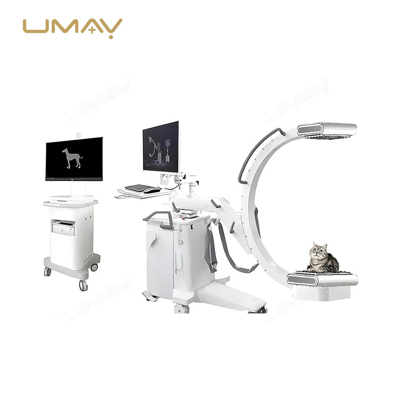
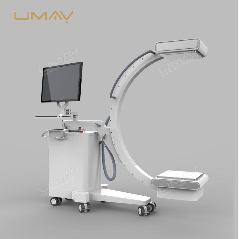
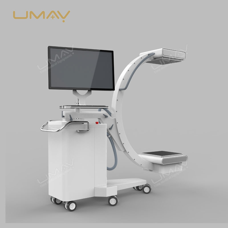
UMARM043 5kW Mobile C-Arm System with Digital Flat Panel Imaging
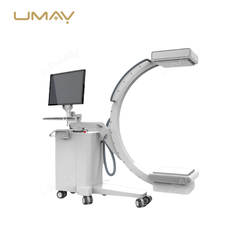
The UMARM043 5kW Mobile C-Arm X-Ray System is designed for fluoroscopic and radiographic imaging in hospitals and medical facilities. The system combines a 5kW high-frequency X-ray generator, digital flat panel detector and mobile C-arm structure to support diagnostic and interventional imaging workflows.
The 210 × 210 mm amorphous silicon TFT detector features a 1024 × 1024 pixel matrix, 200 μm pixel pitch, 16-bit grayscale acquisition and a specified frame rate of 30 fps. The system supports both continuous and pulsed fluoroscopy, together with radiographic image acquisition.
Motorized vertical positioning, manually adjustable C-arm movements, digital image processing and a DICOM 3.0 workstation provide the positioning and image-management functions described in the UMARM043 technical catalog.
Key Features
5kW High-Frequency Generator
Maximum output power of 5 kW, with specified exposure ranges for radiography and fluoroscopy.
210 × 210 mm Flat Panel Detector
Amorphous silicon TFT detector with a 1024 × 1024 acquisition matrix, 200 μm pixel pitch and 16-bit grayscale imaging.
Continuous and Pulsed Fluoroscopy
Supports continuous fluoroscopy and pulsed fluoroscopy with an adjustable pulsed frame rate of 1–30 fps.
Adjustable C-Arm Positioning
Motorized vertical movement and manually controlled horizontal, rotational, angular and orbital adjustments.
Digital Image Processing
Provides image enhancement, window-level adjustment, image rotation, magnification, annotations and measurement tools.
DICOM 3.0 Workstation
Includes a DICOM 3.0 interface, patient information management and digital image archiving functions.
Digital Flat Panel Detector
The UMARM043 is equipped with a monolithic amorphous silicon thin-film transistor (TFT) detector for digital X-ray image acquisition.
| Parameter | Specification |
|---|---|
| Detector Type | Monolithic amorphous silicon TFT |
| Detector Panel Size | 210 × 210 mm |
| Acquisition Pixel Matrix | 1024 × 1024 pixels |
| Pixel Pitch | 200 μm |
| Spatial Resolution | 2.5 LP/mm |
| Pixel Bit Depth | 16-bit grayscale |
| Frame Rate | 30 fps |
5kW High-Frequency X-Ray Generator
The high-frequency generator provides exposure settings for radiographic image acquisition and fluoroscopic examination. The system includes continuous and pulsed fluoroscopy modes and an integrated diagnostic self-test function.
| Parameter | Specification |
|---|---|
| Maximum Output Power | 5 kW |
| Input Power Supply | 220 VAC |
| Radiography Voltage | 40–125 kV |
| Radiography Tube Current | 10–100 mA |
| Exposure Time | 1–5000 ms |
| mAs Range | 0.1–200 mAs |
| Fluoroscopy Voltage | 40–120 kV |
| Continuous Fluoroscopy Current | 0.5–5 mA |
| Pulsed Fluoroscopy Current | 10–30 mA |
| Pulsed Fluoroscopy Frame Rate | 1–30 fps |
| Diagnostic Self-Test | Integrated self-test and display |
X-Ray Tube Assembly
The UMARM043 X-ray tube assembly features dual focal spot sizes of 0.3 mm and 0.6 mm, with a maximum tube voltage of 125 kV.
| Parameter | Specification |
|---|---|
| Focal Spot Sizes | 0.3 mm / 0.6 mm |
| Maximum Tube Voltage | 125 kV |
| Anode Target Angle | 12° |
C-Arm Mechanical System
The UMARM043 combines motorized vertical displacement with manually operated C-arm movements. These adjustments allow operators to position the imaging components according to the required projection and examination setup.
| Mechanical Parameter | Specification |
|---|---|
| Vertical Displacement | Motorized, ≥400 mm |
| Horizontal Displacement | Manual, ≥200 mm |
| Rotational Displacement | Manual, ≥±200° |
| Horizontal Angulation | Manual, ≥±12.5° |
| Orbital Movement | Manual, −30° ±5° to +90° ±5° |
| Depth of Arc | ≥650 mm |
| Source-to-Image Distance (SID) | ≥1000 mm |
Grid and Collimator
The system configuration includes an aluminum-based grid and a motorized DR beam-limiting device.
| Parameter | Specification |
|---|---|
| Grid Material | Aluminum-based grid |
| Grid Size | 23 × 23 cm |
| Grid Ratio | 100:1 |
| Grid Frequency | 215 lines per inch (LPI) |
| Grid Focal Length | 1000 mm |
| Collimator Type | Motorized DR beam-limiting device |
Image Acquisition and Processing
The UMARM043 imaging workstation provides digital image acquisition, image processing, annotation and measurement functions. It also includes patient information management and image archiving capabilities.
Window and Level Control
Adjustable and preset window width and level, including local automatic window-level adjustment.
Image Display Tools
Image inversion, rotation, magnification, panning and localized image enlargement.
Image Enhancement
Edge enhancement, interpolation, image correction, sharpening, filtering and image-restoration functions.
Annotation and Measurement
Text, number and marker annotations, with line, angle, area and arbitrary-shape measurement tools.
Acquisition and Processing Tools
Image stitching, automatic exposure index display and customizable acquisition protocols for anatomical regions.
Patient Data Management
Integrated patient data management and image archiving with a DICOM 3.0 interface.
Diagnostic Workstation Specifications
| Component | Specification |
|---|---|
| Processor | Intel Core i7, 3 GHz or higher |
| Memory | ≥16 GB DDR3, 1600 MHz |
| Storage | 1 TB, 7200 RPM hard drive |
| Display | IPS LCD monitor |
| Display Resolution | 1920 × 1080 (Full HD) |
| Network Connectivity | 1000 Mbps Ethernet, Jumbo Frames up to 9 KB |
| DICOM Interface | DICOM 3.0 |
| User Interface Language | Chinese language support |
| Operating System | Windows 10 64-bit, Professional Edition or higher |
Diagnostic and Interventional Imaging Applications
The UMARM043 is described in the product catalog as a mobile C-arm system for diagnostic and interventional imaging procedures. Its fluoroscopy and radiography modes provide digital image acquisition functions for relevant clinical workflows.
Continuous Fluoroscopy
Continuous fluoroscopic acquisition within the specified 40–120 kV voltage and 0.5–5 mA current ranges.
Pulsed Fluoroscopy
Pulsed fluoroscopic acquisition with a specified frame-rate range of 1–30 fps.
Radiographic Imaging
Radiographic image acquisition using the 5kW generator and its specified radiography exposure settings.
Mobile C-Arm Positioning
Wheeled equipment configuration and adjustable C-arm geometry support positioning around the examination area.
Before Ordering
Please provide the following information so UMY Medical can review the appropriate UMARM043 configuration and prepare a quotation.
- Required quantity and destination country.
- Intended diagnostic or interventional imaging application.
- Required fluoroscopy and radiography functions.
- Flat panel detector and monitor requirements.
- Available room space and C-arm movement requirements.
- Local power supply and installation conditions.
- Required software language and DICOM integration.
- Workstation and accessory requirements.
- Required technical documents and local regulatory information.
- Delivery, installation, training and service requirements.
Request a Quote for UMARM043
Contact UMY Medical for the UMARM043 5kW Mobile C-Arm X-Ray System, including detailed technical specifications, configuration information and export quotation.
Please share your required quantity, destination country, intended imaging application and any specific project requirements.
Frequently Asked Questions
What type of detector does the UMARM043 use?
The UMARM043 is specified with a 210 × 210 mm amorphous silicon TFT flat panel detector featuring a 1024 × 1024 pixel acquisition matrix, 200 μm pixel pitch and 16-bit grayscale depth.
Does the UMARM043 support continuous and pulsed fluoroscopy?
Yes. The 5kW generator supports continuous fluoroscopy at 0.5–5 mA and pulsed fluoroscopy at 10–30 mA, with a specified pulsed frame-rate range of 1–30 fps.
What positioning functions does the C-arm provide?
The system includes motorized vertical displacement of at least 400 mm, together with manual horizontal displacement, rotational adjustment, horizontal angulation and orbital movement.
Does the UMARM043 workstation support DICOM?
Yes. The product catalog specifies a DICOM 3.0 workstation interface, integrated patient data management and image archiving. Specific integration requirements should be included in the purchasing inquiry.