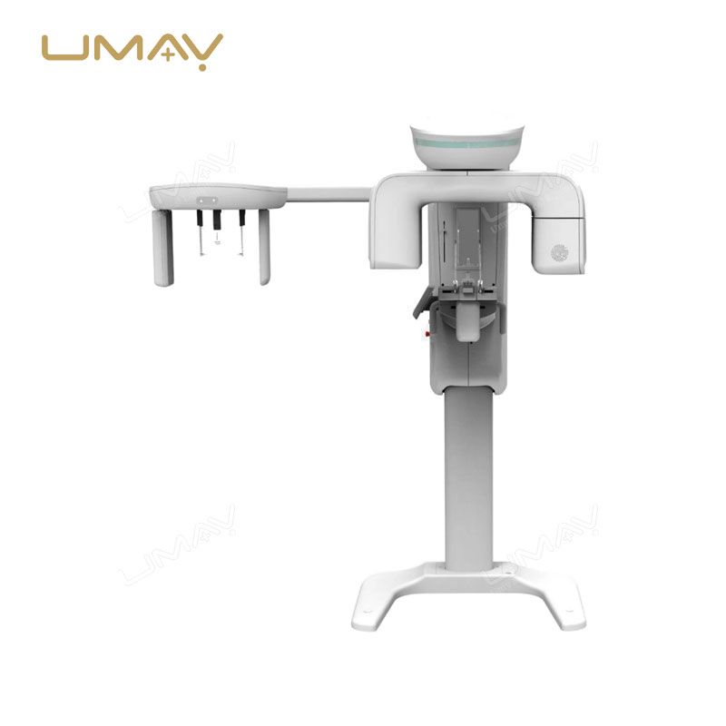
Italy IMD Brand,fixed anode, dual focus 0.6/1.5mm
| ITEMS | PARAMETERS |
|---|---|
| HF generator | 120kV,5kW ,40kHz |
| Fluoroscopy mode 40~120kV 0.5~5mA | |
| (KV manual and automatic); | |
| Fluoroscopy mode | Pulsd fluoroscopy 40~120kV 0,5-5mA |
| Boost fluoroscopy 40~120kV 5~10mA; | |
| Photography 40~120kV 1~250mAs | |
| X- ray tube | Dual-focus Rotating anode: 0.3mm/0.6mm |
| Image intensifier | Three fields of vision 9, 6, 4.5 French Thales Brand |
| CCD camera | CCD camera, Japanese Watt Brand, 360° rotation |
| Recursive Noise Reduction | |
| Frame freezing | |
| Image system | Multi-frame image storage |
| Image conversing and rotating | |
| Screen display with image comparison | |
| Mechanical motion specifications | Verticaltravel: 0-400mm |
| Horizontaltravel: 0~200mm | |
| Rotation about horizontal axis: ±180° | |
| Distance of focal spot to image generator(SID): 1000mm | |
| Depth in arm≥650mm | |
| Mobile stand : 1800mm*800mm*1850mm | |
| C-arm dimension | Image system: 750mm*530mm*1680mm |
| High resolution CCD camera: 1set | |
| x-ray generator: 1set | |
| 9” image intensifier: 1set | |
| Standard configuration | image processing system: 1set |
| LCD high definition display: 2 sets | |
| Mobile stand : 1set | |
| ITEMS | PARAMETERS |
|---|---|
| Application | x ray machine |
| Function | One step to control dental x ray expose |
| LED | No |
| Current | 3A |
| Housing materials | ABS plastic |
| Operation voltage | 125v |
| Standard core | 2 core |
| Standard Length | 6m wire (60cm spring coil wire) |
| Mechanical life | ≤200,000 times |
| Electrical life | ≤400,000 times |
| Structure | omron inner switch |
| Customization | Available |
| Power Source | Manual |
| Instrument classification | Class I |
Specific Parameters
| ITEMS | PARAMETERS | REMARKS |
|---|---|---|
| X-ray Generator | Generator Type: High Frequency Inverter 80kHz | Self-developed and world advanced all-solid-state high frequency high voltage x-ray generator |
| Input Power: Single phase 220VAC, 50/60Hz | ||
| Radiographic Ratings: | ||
| Large Focal Point 20-35kV/10-510mAs | ||
| Small Focal Point 20-35kV/10-100mAs | ||
| Power Rating: 6.2kVA | ||
| X-ray Tube | Focal Spot Size: Dual Focus 0.1 / 0.3mm | Model: IAE TUBE FROM ITALY |
| Target Material: Molybdenum (Mo) | ||
| Port Material: Beryllium (Be) | ||
| High-speed anode drive: 2800 /10000rpm | ||
| Target angle: 10°/16° | ||
| Anode Heat Storage: 210kJ (300kHU) | ||
| Anode Cooling: Air cooling | ||
| Filtration: Mo(0.03mm), Al(0.5mm) | ||
| Radiographic Stand | C-ARM: Vertical Movement: 590mm | Electric Isocentric rotating |
| Center of electric rotating C-arm | ||
| Automatic return function by one key | ||
| Rotations Degree: +90°~-90° | ||
| Automatically released after the exposure pressure settings display | ||
| Compression flexible, stepless speed. | ||
| Max. pressure: 200N | ||
| Max. travel: 150mm | ||
| SID: 650mm | ||
| Flat Panel Detector | Detector material: Amorphous silicon | China Flat Panel Detector |
| Effective coverage of detector: 24x30cm | ||
| Pixel matrix: 3072×1944 | as standard | |
| Limit of spatial resolution: 6.0Lp/mm | ||
| DQE value: 70% | ||
| dynamic range: 14bit digital output | ||
| pixel size: 75μm | ||
| High voltage Synchronizer trigger: BNC | ||
| Output: Camera Link or Ethernet | ||
| Working condition: 10ºC-40ºC | ||
| storage environment: -10ºC-50ºC | ||
| Bucky housing and movement device | Size: 374*304*65mm | |
| Stepless speed regulating range: 0~6cm/s | ||
| Movement range: 0.5~2cm | ||
| Grid Size: 24x30cm | ||
| Grid ratio: 5:1 | ||
| Grid density: 30lp/cm | ||
| Focal distance: 650mm | ||
| Image acquisition workstation | CPU≥Intel Core Duo 2.60GHz | Configuration including Diagnose digital workstation |
| Hardware≥250G high speed Hardware | ||
| Memory≥2G | 5M medical monitor for optional | |
| Display card≥512MB | ||
| high brightness high-contrast LCD,1280*1024 Pixel resolution | ||
| Network interface Work-list | ||
| DICOM3.0 transmission | ||
| 100/1000 Gigabit Ethernet | ||
| Software:Imaging software package DMOC V1.0 | ||
| Others | Line Voltage | 110V for optional |
| 220V ac±10% 25A,Single phase | ||
Product Details
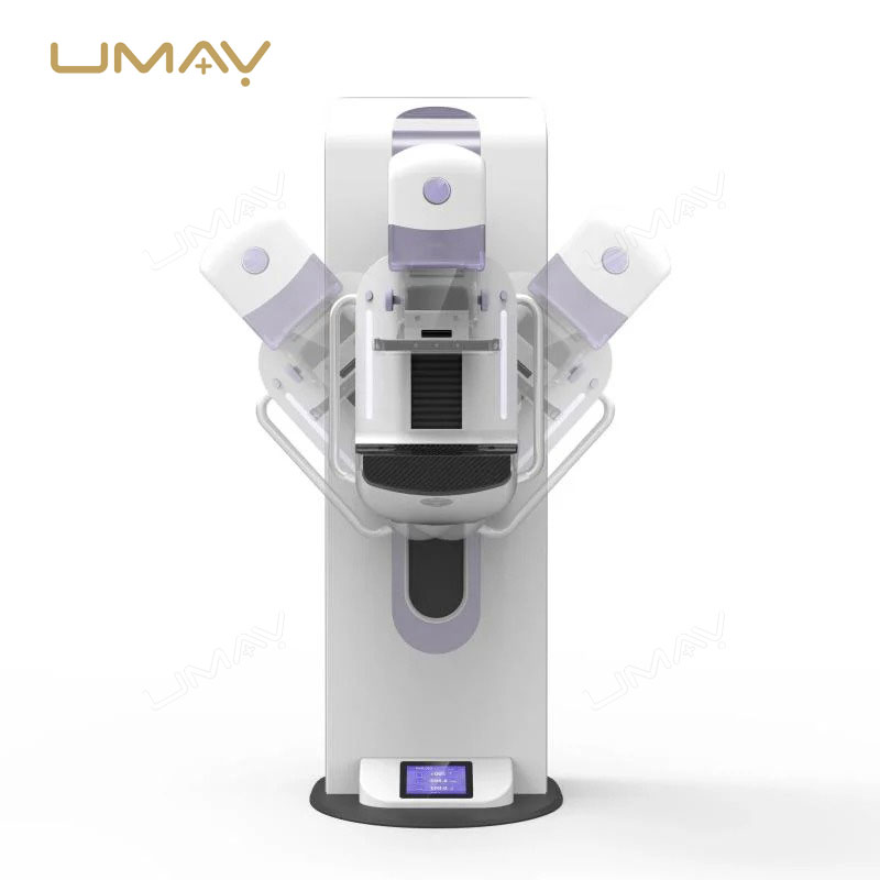
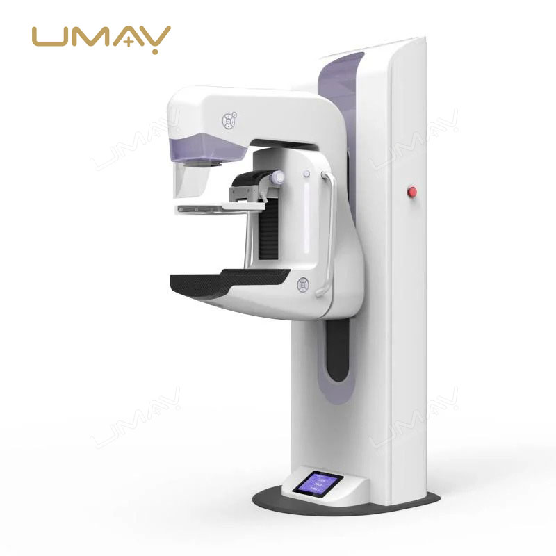
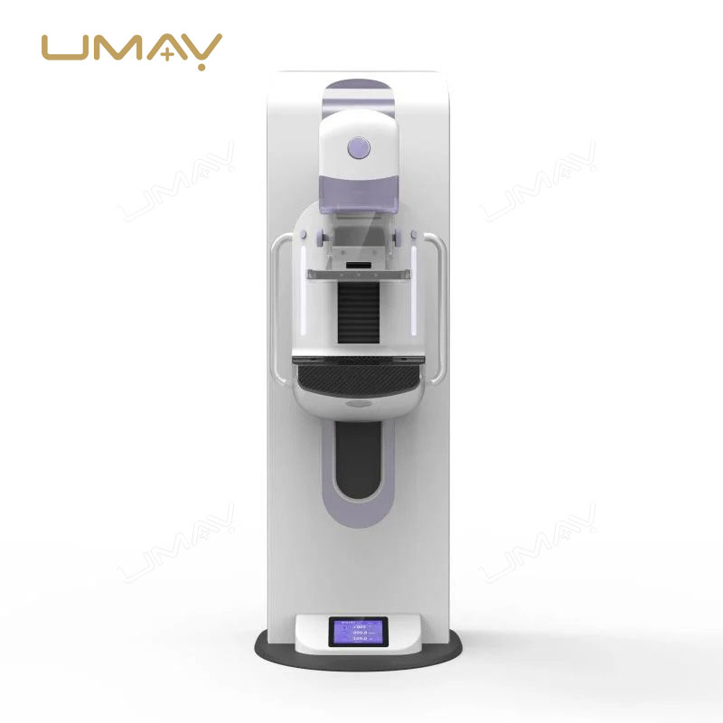
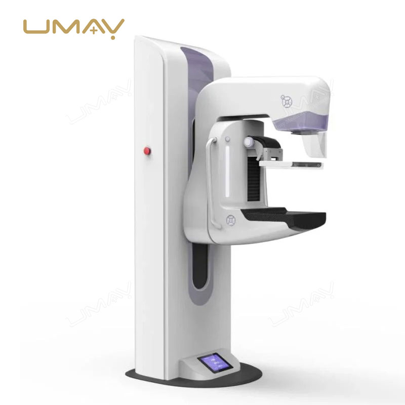
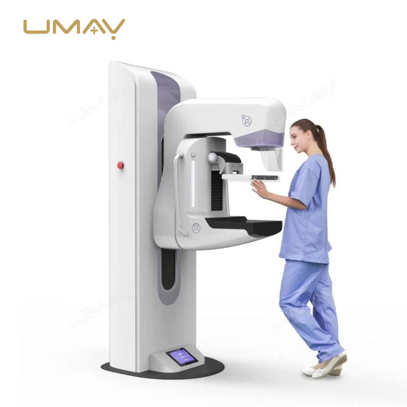
- UMY-XM-MA02 Enhanced Breast Mammography X-Ray Machine: Advanced digital mammography system designed for accurate breast cancer screening and diagnosis.
- High-Resolution Breast Imaging: Produces exceptionally detailed images for precise evaluation of breast tissue and abnormalities.
- Optimized for Early Breast Cancer Detection: Supports identification of suspicious lesions at the earliest possible stage.
- Advanced Digital Mammography Technology: Delivers superior image quality with fast image acquisition and processing.
- Enhanced Diagnostic Accuracy: Provides clear visualization of breast structures for confident clinical assessment.
- Excellent Microcalcification Detection: Captures fine calcifications that may indicate early signs of breast disease.
- Superior Soft Tissue Contrast: Improves differentiation between normal and abnormal breast tissue.
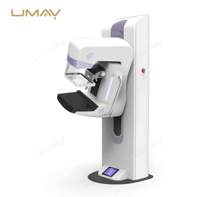
- Low Radiation Dose Operation: Minimizes patient exposure while maintaining exceptional image quality.
- Precise Tumor Identification: Helps detect small masses and subtle tissue changes that may be difficult to identify with conventional imaging.
- Reliable Breast Screening Solution: Ideal for routine breast examinations and preventive healthcare programs.
- Fast Image Acquisition: Reduces examination time and improves patient throughput in busy clinical environments.
- Advanced Image Processing: Enhances image sharpness, contrast, and diagnostic confidence.
- Supports Comprehensive Breast Assessment: Enables detailed evaluation of breast anatomy and pathological findings.
- User-Friendly Operating Interface: Simplifies workflow and improves productivity for radiographers and clinicians.
- Efficient Clinical Workflow: Streamlines breast imaging procedures and accelerates diagnostic reporting.
- Consistent Imaging Performance: Ensures stable, reproducible image quality across screening and diagnostic examinations.
- Digital Data Management Compatibility: Integrates seamlessly with PACS, RIS, and hospital information systems.
Detail Gallery