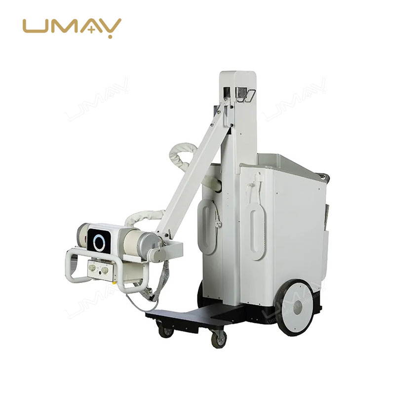
Italy IMD Brand,fixed anode, dual focus 0.6/1.5mm
| ITEMS | PARAMETERS |
|---|---|
| HF generator | 120kV,5kW ,40kHz |
| Fluoroscopy mode 40~120kV 0.5~5mA | |
| (KV manual and automatic); | |
| Fluoroscopy mode | Pulsd fluoroscopy 40~120kV 0,5-5mA |
| Boost fluoroscopy 40~120kV 5~10mA; | |
| Photography 40~120kV 1~250mAs | |
| X- ray tube | Dual-focus Rotating anode: 0.3mm/0.6mm |
| Image intensifier | Three fields of vision 9, 6, 4.5 French Thales Brand |
| CCD camera | CCD camera, Japanese Watt Brand, 360° rotation |
| Recursive Noise Reduction | |
| Frame freezing | |
| Image system | Multi-frame image storage |
| Image conversing and rotating | |
| Screen display with image comparison | |
| Mechanical motion specifications | Verticaltravel: 0-400mm |
| Horizontaltravel: 0~200mm | |
| Rotation about horizontal axis: ±180° | |
| Distance of focal spot to image generator(SID): 1000mm | |
| Depth in arm≥650mm | |
| Mobile stand : 1800mm*800mm*1850mm | |
| C-arm dimension | Image system: 750mm*530mm*1680mm |
| High resolution CCD camera: 1set | |
| x-ray generator: 1set | |
| 9” image intensifier: 1set | |
| Standard configuration | image processing system: 1set |
| LCD high definition display: 2 sets | |
| Mobile stand : 1set | |
| ITEMS | PARAMETERS |
|---|---|
| Application | x ray machine |
| Function | One step to control dental x ray expose |
| LED | No |
| Current | 3A |
| Housing materials | ABS plastic |
| Operation voltage | 125v |
| Standard core | 2 core |
| Standard Length | 6m wire (60cm spring coil wire) |
| Mechanical life | ≤200,000 times |
| Electrical life | ≤400,000 times |
| Structure | omron inner switch |
| Customization | Available |
| Power Source | Manual |
| Instrument classification | Class I |
Specific Parameters
| ITEMS | PARAMETERS |
|---|---|
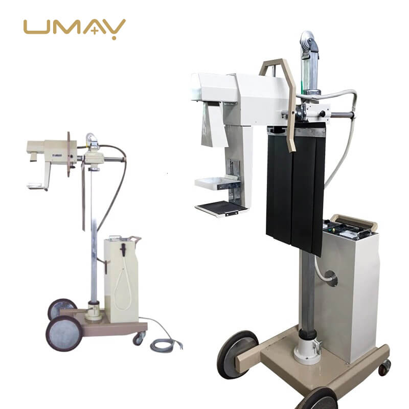
| System Overview (Basic Requirement) | Clinical application: dedicated to human breast tissue X-ray photography to obtain tissue images for breast disease diagnosis and breast cancer screening; |
| Frame Structure | Rack structure: The C arm is developed and produced by manufacturer. (Possess all independent intellectual property rights) |
| C arm rotation range: -155°~ +195° | |
| C arm vertical movement range: 630 ~ 1440mm; | |
| C arm can be locked anywhere in the full stroke | |
| The display on the rack can display the current position and the position information to be photographed, and has a one-key switch function to the preset position; | |
| FID: 660mm; | |
| High Voltage Generator Component | Maximum output power: 5kW |
| Output voltage range: 20 ~ 40 kV | |
| Output current range: 10 ~ 160 mA | |
| Loading time range: 0.005s ~ 10s | |
| Current time product range: 0.5 ~ 560mAs; | |
| With exposure control system, support for one-key exposure or hand brake exposure | |
| X-ray Tube Component | Double speed rotating anode |
| Nominal tube voltage: 40 KV; | |
| Anode heat capacity: 300 kHU; | |
| Anode rotating speed: 10000 rpm | |
| Anode material: molybdenum | |
| Nominal focus size: small focus 0.1mm, large focus 0.3mm; | |
| Flat Panel Detector Component | Imaging material: amorphous silicon |
| Imaging size: 240 × 300mm; | |
| Conventional spatial resolution: 7.01 p/mm; | |
| Pixel matrix: 2816 × 3584mm pixels; | |
| A/D output: 16 bits; | |
| Cooling method: fan cooling | |
| Weight: 3.5 kg | |
| Pixel size: 85 μm | |
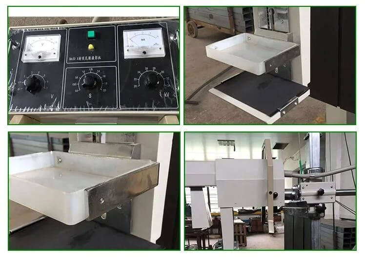
Product Details
- Digital High-Performance Mammography Machine: Advanced breast imaging system designed for accurate screening and diagnostic mammography.
- High-Resolution Digital Imaging: Produces exceptionally clear and detailed breast images for confident clinical evaluation.
- Optimized for Breast Cancer Screening: Supports early detection of breast cancer and other breast abnormalities.
- Advanced Digital Mammography Technology: Delivers superior image quality with fast acquisition and processing speeds.
- Exceptional Tissue Visualization: Enhances visibility of breast structures, masses, and subtle abnormalities.
- High Diagnostic Accuracy: Provides precise imaging to support reliable diagnosis and treatment planning.
- Low Radiation Dose Operation: Minimizes patient exposure while maintaining outstanding image clarity.
- Excellent Microcalcification Detection: Captures fine calcifications that may indicate early-stage breast disease.
- Enhanced Soft Tissue Contrast: Improves differentiation of dense and soft breast tissues.
- Fast Image Acquisition: Reduces examination times and increases patient throughput.
- Efficient Workflow Management: Streamlines imaging procedures for busy hospitals and breast imaging centers.
- User-Friendly Operating Interface: Simplifies operation and improves productivity for technologists and radiologists.
- Reliable Clinical Performance: Ensures consistent image quality across routine screening and diagnostic examinations.
- Supports High-Volume Screening Programs: Ideal for hospitals and healthcare facilities with large patient volumes.
- Advanced Image Processing Algorithms: Enhance image sharpness, contrast, and diagnostic confidence.
- Comfort-Oriented Examination Design: Helps improve the patient experience during breast imaging procedures.
- Digital Data Management Compatibility: Integrates seamlessly with PACS, RIS, and hospital information systems.
- Rapid Image Review: Allows healthcare professionals to access and evaluate images immediately after acquisition.
- Durable Medical-Grade Construction: Built for long-term reliability and continuous clinical operation.
- Enhanced Radiologist Confidence: Provides detailed imaging for accurate interpretation and informed decision-making.
- Supports Preventive Healthcare Programs: Plays a critical role in routine breast health screening initiatives.
- Improved Patient Safety: Combines advanced imaging performance with optimized low-dose technology.
- Suitable for Screening and Diagnostic Applications: Effective for routine examinations, follow-up studies, and comprehensive breast assessments.
- Ideal for Hospitals and Imaging Centers: Designed to meet the demands of modern breast care and radiology departments.
Detail Gallery