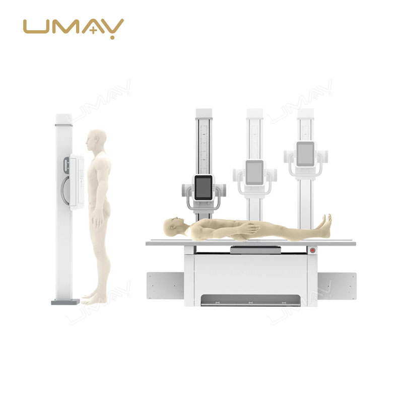
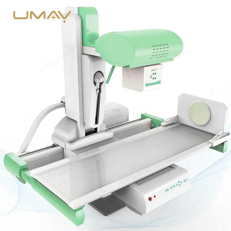
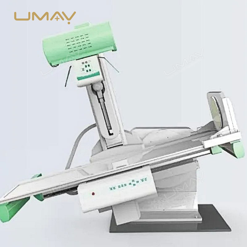
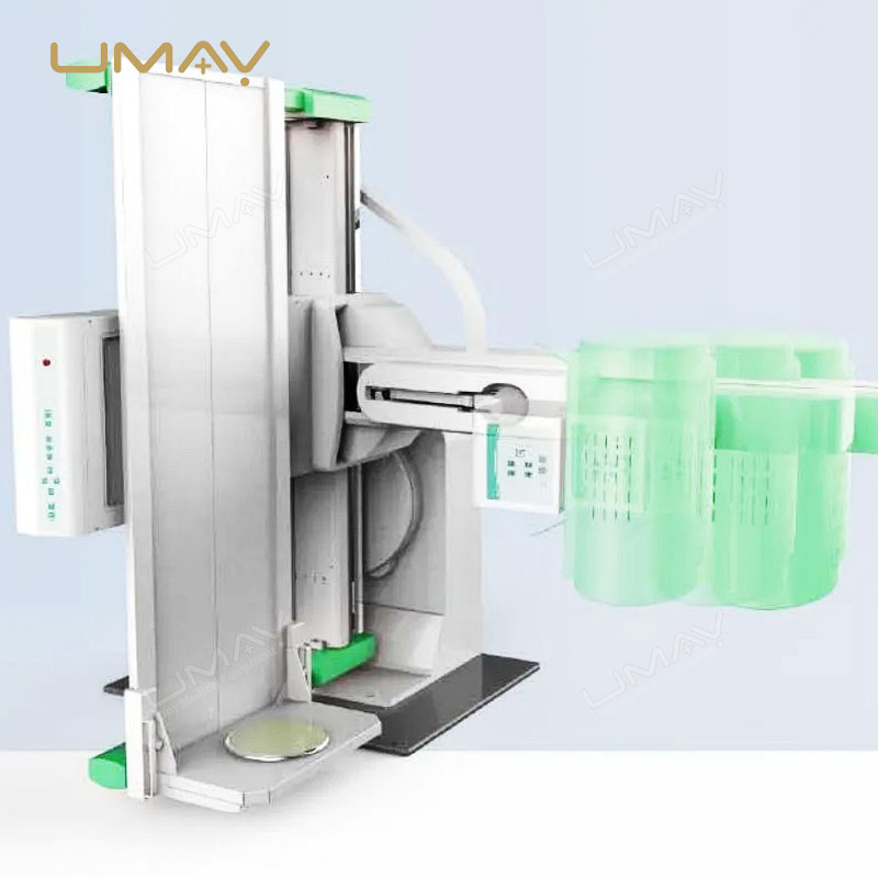
The Digital Radiography and Fluoroscopy (DRF) System offers advanced imaging technology for high-precision diagnostics. Combining the benefits of digital radiography with real-time fluoroscopy, this system ensures clear, detailed images for a wide range of applications, including orthopedics, cardiology, and gastrointestinal procedures. It provides accurate, high-resolution images with minimal radiation exposure, enhancing both patient safety and diagnostic efficiency. Ideal for hospitals and specialized imaging centers, this DRF system supports rapid decision-making and precise treatment planning. Its advanced features make it an indispensable tool for healthcare professionals seeking superior imaging capabilities in complex diagnostic procedures.
Specific Parameters
| ITEMS | PARAMETERS |
|---|---|
| Power Supply | |
| Voltage | 380V ± 38V |
| Frequency | 50Hz ± 1Hz |
| Capacity | ≥65kVA |
| Internal Resistance | ≤0.17Ω |
| Generator | |
| Power | 80kW |
| Inverter Frequency | 440kHz |
| Radiography Tube Voltage | 40kV ~ 150kV step adjustment |
| Radiography Tube Current | 10mA ~ 1000mA step adjustment |
| Radiography Time | 1.0ms ~ 10,000ms step adjustment |
| Radiography mAs | 0.1mAs ~ 1000mAs |
| Fluoroscopy Tube Voltage | 40kV ~ 125kV step adjustment |
| Fluoroscopy Tube Current | 0.5mA ~ 10mA (continuous fluoroscopy), 5mA ~ 20mA (pulse fluoroscopy) |
| X-Ray Tube Assembly | |
| Target | Molybdenum-based lanthanum-tungsten composite |
| Target Angle | 12° |
| Nominal Tube Voltage | 150kV |
| Tube Focus (Big/Small) | 1.2mm / 0.6mm |
| Input Power | Big Focus: 100kW, Small Focus: 40kW |
| Anode Thermal Capacity | 420kJ (600kHU) |
| Anode Maximum Heat Dissipation | 1750W (2465kHU/min) |
| Component Heat Capacity | 1420kJ (2000kHU) |
| Anode Rotation Speed | 9700rpm |
| Collimator | |
| Field of View Light | Halogen lamp, AC24V/100W |
| Visible Light Illumination | Average illumination brightness > 100LUX |
| Light Field Exposure Time | 5 ~ 45s, 5s per step |
| Diagnostic Table | |
| Table Transverse Movement Distance | 300mm |
| SID | 1100mm ~ 1800mm |
| Table Panel Equivalent Filtration | ≤1.2mmAl |
| Point Device Movement Range | 1250mm |
| Table Height from Ground | 905mm |
| Table Size | 2100mm × 730mm |
| Table Rotation | +90° ~ -25° |
| X-Ray Tube Column Swing | +40° ~ 0° ~ -40° |
| Minimum Distance of Compressor from Table Surface | ≤150mm |
| Compressor Pressure | 80N ~ 130N |
| Table Bearing | 135kg |
| Fragmentation | Whole film, two-piece, four-slice |
| Fixed Grid | |
| Grid Density | 103L/INCH |
| Ratio | 0.41736111111111 |
| Convergence Distance | 130cm |
| Stationary Type | 15″ × 18″ |
| Intensifier | |
| Field of View Size | 230mm |
| Resolution | 52 Lp/cm |
| Conversion Factor | 26 |
| Signal-to-Noise Ratio | 45 dB |
| X-Ray Camera | |
| Cell Size | 5.86 × 5.86µm |
| Number of Valid Pixels | 1920 × 1200 |
| Flat Panel Detector | |
| Image Sensor | Amorphous silicon thin-film transistor |
| Pixel Size | 143µm |
| Effective Pixel Size | 3008(H) × 3072(V) |
| Effective Area (H × V) | 430(H) × 439(V) |
| Gray Scale | 14-bit |
| Spatial Resolution | 3.7 Lp/mm (CsI) |
| Energy Range | 40kV ~ 150kVP |
| Power Input | DC24V |
Product Details
- Digital Radiography and Fluoroscopy (DRF) System: Combines digital radiography and real-time fluoroscopy technology for advanced diagnostic imaging applications.
- High-Precision Imaging Performance: Provides clear, detailed, and high-resolution images for accurate medical diagnosis and treatment planning.
- Real-Time Fluoroscopy Capability: Supports continuous live imaging for dynamic examinations and interventional procedures.
- Advanced Digital Radiography Technology: Delivers fast image acquisition with enhanced image clarity and diagnostic accuracy.
- Minimal Radiation Exposure: Optimized imaging technology helps reduce radiation dose while maintaining excellent image quality.
- Ideal for Orthopedic Applications: Supports bone, joint, and musculoskeletal examinations with precise radiographic visualization.
- Suitable for Cardiology Procedures: Provides reliable imaging guidance for cardiovascular diagnostics and interventional procedures.
- Optimized for Gastrointestinal Examinations: Enables dynamic fluoroscopic imaging for digestive system evaluation and contrast studies.
- High-Resolution Diagnostic Imaging: Enhances visualization of anatomical structures and pathological conditions for improved clinical confidence.
- Rapid Decision-Making Support: Real-time imaging and fast processing assist healthcare professionals in making timely clinical decisions.
- User-Friendly Operation Interface: Intuitive control system simplifies examination setup, image acquisition, and workflow management.
- Efficient Clinical Workflow: Streamlines imaging procedures and improves productivity in hospitals and imaging centers.
- Versatile Multi-Department Applications: Suitable for radiology, orthopedics, cardiology, gastroenterology, emergency medicine, and surgical departments.
- Advanced Image Processing Technology: Provides enhanced image optimization, contrast adjustment, and diagnostic analysis capabilities.
- Reliable and Stable System Performance: High-quality components ensure consistent imaging performance and long-term operational reliability.
- Durable Medical-Grade Construction: Designed for continuous daily use in demanding healthcare environments.
- Enhanced Patient Safety: Low-dose imaging technology supports safer examinations without compromising diagnostic quality.
- Supports Precise Treatment Planning: Accurate imaging assists physicians in diagnosis, intervention guidance, and treatment evaluation.
- Ideal for Hospitals and Specialized Imaging Centers: Perfect for advanced diagnostic facilities requiring high-performance DR and fluoroscopy capabilities.
- Comprehensive Medical Imaging Solution: An essential system for healthcare professionals seeking reliable, efficient, and superior imaging performance in complex diagnostic procedures.
Detail Gallery