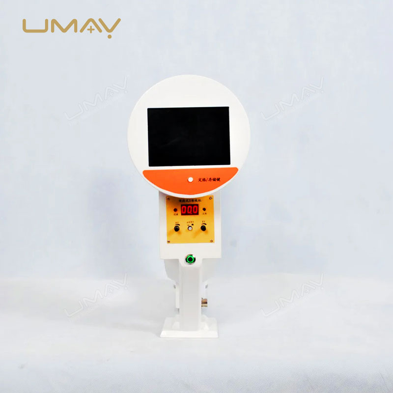


Mobile C-Arm Fluoroscopy for Surgical Imaging
The 3.5kW Mobile C-Arm X-Ray System is designed for real-time fluoroscopic imaging during orthopedic, trauma, spinal, urological and general surgical procedures. Its mobile C-arm structure supports flexible positioning around the operating table for intraoperative X-ray examination.
The system combines a 3.5kW high-frequency X-ray generator, a 9-inch image intensifier and a high-resolution CCD camera. Continuous, pulsed and boost fluoroscopy modes provide different imaging options, while digital noise reduction, last-image hold and image saving functions support intraoperative image review.
With a 10.4-inch LCD control interface, adjustable mechanical positioning and a 15-inch display monitor, the system is suitable for surgical departments and clinical environments requiring mobile fluoroscopic imaging.
Key Features
3.5kW High-Frequency Generator
A 40kHz inverter generator supports X-ray operation at 40–110kV for fluoroscopic imaging and radiographic exposure.
9-Inch Image Intensifier
Provides three selectable fields of view: 9 inches, 6 inches and 4.5 inches, supporting different imaging areas and magnification requirements.
Multiple Fluoroscopy Modes
Supports continuous, pulsed and boost fluoroscopy with adjustable exposure parameters for different intraoperative imaging requirements.
High-Resolution CCD Imaging
A CCD camera with 360° rotation works with the image intensifier to acquire fluoroscopic images for display and review.
Digital Image Processing
Includes digital noise reduction, last-image hold, image saving and image inversion functions to support image examination during procedures.
Adjustable Mobile C-Arm
Vertical travel, horizontal movement, C-arm rotation and a movable stand provide positioning flexibility around surgical tables.
Fluoroscopy and Imaging Functions
The 3.5kW Mobile C-Arm X-Ray System provides multiple operating modes for surgical fluoroscopy. The high-frequency generator and image intensifier-based imaging system support real-time visualization during selected clinical procedures.
Continuous Fluoroscopy
Supports continuous imaging at 40–110kV and 0.5–5mA, with manual and automatic kV control described in the technical documentation.
Pulsed Fluoroscopy
Provides pulsed image acquisition at 40–110kV and 0.5–5mA. Actual radiation exposure depends on the selected operating parameters and examination conditions.
Boost Fluoroscopy
Offers a 40–110kV operating range with 5–10mA tube current for the additional fluoroscopy mode specified by the manufacturer.
Image Storage and Review
Last-image hold, image saving, noise reduction and image inversion support review of acquired fluoroscopic images.
Fluoroscopy and Radiography Parameters
The system provides the following operating modes and exposure ranges. Clinical users should select appropriate settings according to the intended procedure, patient characteristics and radiation protection requirements.
| Operating Mode | Voltage Range | Current / Exposure Range |
|---|---|---|
| Continuous Fluoroscopy | 40–110kV | 0.5–5mA |
| Pulsed Fluoroscopy | 40–110kV | 0.5–5mA |
| Boost Fluoroscopy | 40–110kV | 5–10mA |
| Radiography | 40–110kV | 1–250mAs |
- Manual and automatic kV adjustment are listed for the fluoroscopy mode.
- Pulsed fluoroscopy provides an alternative to continuous exposure according to the selected clinical workflow.
- Actual radiation dose depends on imaging settings, patient characteristics and operating technique.
- Specific dose reduction percentages are not established by the supplied technical specifications.
Technical Specifications
| Specification | Technical Details |
|---|---|
| Product Type | High-Frequency Mobile C-Arm X-Ray System |
| Generator Power | 3.5kW |
| Generator Frequency | 40kHz |
| X-Ray Tube Voltage | 40–110kV |
| Maximum Tube Voltage | 110kV |
| X-Ray Tube Type | Fixed anode, dual focal spot |
| Focal Spot Sizes | 0.6mm / 1.5mm |
| Imaging Technology | Image intensifier with CCD camera |
| Image Intensifier | 9 inches |
| Selectable Fields of View | 9″ / 6″ / 4.5″ |
| Imaging Camera | High-resolution CCD camera |
| CCD Camera Rotation | 360° |
| Control Interface | 10.4-inch LCD |
| Control Panel Rotation | 180° total (90° left / 90° right) |
| Display Monitor | 15-inch high-resolution monitor |
| Image Processing | Digital noise reduction, last-image hold, image saving and image inversion |
| Equipment Structure | Mobile C-arm with adjustable positioning |
- The specifications are based on the supplied 3.5kW mobile C-arm technical documentation.
- The standard imaging system uses an image intensifier and CCD camera rather than a flat panel detector.
- Detector alternatives, DICOM/PACS interfaces and advanced angiography software are not established by the supplied specifications.
- Final component brands, software functions and accessories should be confirmed before quotation.
C-Arm Movement and Mechanical Specifications
The mobile C-arm provides adjustable positioning for fluoroscopic imaging around the patient. Vertical and horizontal movement, rotational adjustment and a mobile stand help accommodate different operating table arrangements.
| Mechanical Parameter | Specification |
|---|---|
| Vertical Travel | 0–400mm |
| Horizontal Travel | 0–200mm |
| C-Arm Rotation Angle | ≥115° |
| Lateral Angulation | ≥±12.5° |
| Source-to-Image Distance | 1000mm |
| C-Arm Depth | ≥650mm |
| Mobile Stand Dimensions | 1800 × 800 × 1850mm |
Mobile Positioning
A steering handle supports movement and positioning of the system within the operating environment.
Parallel Movement
The manufacturer-listed parallel movement function provides an additional positioning adjustment.
Wheel Locking Mechanism
The mobile base incorporates a wheel locking mechanism intended to help secure the equipment after positioning.
Adjustable Control Interface
The LCD control panel can rotate to improve access from different operating positions.
Standard System Configuration
The standard configuration includes the major components required for image intensifier-based mobile C-arm fluoroscopy.
| Component | Quantity |
|---|---|
| High-Frequency X-Ray Generator | 1 set |
| 9-Inch Image Intensifier | 1 set |
| High-Resolution CCD Camera | 1 set |
| Digital Noise Reduction System | Included |
| 15-Inch High-Resolution Monitor | 1 set |
| Mobile C-Arm Stand | 1 set |
The final supply list, electrical requirements, operating accessories, software functions and packaging details should be confirmed before ordering.
Clinical Applications
The mobile C-arm is intended for intraoperative X-ray imaging in surgical and clinical departments where fluoroscopic visualization is required.
Orthopedic Surgery
Supports fluoroscopic imaging during fracture reduction, bone positioning and fixation procedures.
Trauma Surgery
Provides intraoperative imaging assistance during selected trauma and skeletal procedures.
Spine Surgery
Supports fluoroscopic visualization during selected spinal procedures requiring X-ray guidance.
Urological Procedures
Provides X-ray visualization for selected urological procedures requiring intraoperative imaging.
General Surgery
Supports real-time fluoroscopic imaging during selected general surgical procedures.
Operating Room Imaging
The mobile design allows positioning within operating rooms and other suitably equipped clinical imaging environments.
The suitability of the equipment for a specific clinical procedure should be assessed according to imaging requirements, operator training and applicable local regulations.
System Configuration and Installation
The 3.5kW Mobile C-Arm X-Ray System combines a high-frequency X-ray generator, image intensifier, CCD camera, digital image processing system, monitor and mobile C-arm stand.
Before installation, hospitals and distributors should review operating room dimensions, equipment movement clearance, electrical supply, radiation shielding and the intended examination workflow.
Specific software connectivity, hospital network integration and destination-market regulatory documentation should be confirmed during technical review. These capabilities should not be assumed from the standard equipment configuration.
Before Ordering
To confirm the appropriate 3.5kW mobile C-arm configuration and prepare an accurate quotation, please provide the following information:
- Required quantity and destination country.
- Intended application: orthopedic, trauma, spine, urology or general surgery.
- Required fluoroscopy modes and imaging functions.
- Available operating room space and positioning requirements.
- Electrical supply and installation requirements.
- Required monitors, accessories and image storage functions.
- Any required software or hospital system integration.
- Operator training and after-sales support requirements.
- Required technical, regulatory or tender documentation.
- Expected delivery schedule and procurement requirements.
Discover more options in our C-Arm X-ray Machine range.
Request a 3.5kW Mobile C-Arm X-Ray System Quote
Contact UMY Medical to discuss the 3.5kW mobile C-arm system, surgical imaging requirements, equipment configuration and export quotation for your hospital or medical equipment project.
Please include your destination country, required quantity and intended clinical application. Our team can review your technical requirements before preparing a quotation.
Frequently Asked Questions
What is the power of this mobile C-arm X-ray system?
The system uses a 3.5kW high-frequency X-ray generator operating at 40kHz. The specified X-ray tube voltage range is 40–110kV.
Does this C-arm use a flat panel detector?
The standard configuration uses a 9-inch image intensifier and a high-resolution CCD camera. A flat panel detector is not listed in the supplied standard configuration.
What fluoroscopy modes are supported?
The system supports continuous, pulsed and boost fluoroscopy, along with radiographic exposure. Operating voltage, current and exposure ranges are provided in the technical specifications.
What information is required before purchasing?
Buyers should confirm the destination country, intended procedures, power supply, available installation space, required software and accessories, technical documentation and after-sales requirements before ordering.